

Increased basal activity is a key determinant in the severity of human skeletal dysplasia caused by TRPV4 mutations
- PMID: 21573172
- PMCID: PMC3088684
- DOI: 10.1371/journal.pone.0019533
Increased basal activity is a key determinant in the severity of human skeletal dysplasia caused by TRPV4 mutations
Abstract
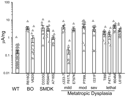
TRPV4 is a mechanically activated Ca(2+)-passing channel implicated in the sensing of forces, including those acting on bones. To date, 33 mutations are known to affect human bone development to different extents. The spectrum of these skeletal dysplasias (SD) ranges from dominantly inherited mild brachylomia (BO) to neonatal lethal forms of metatropic dysplasia (MD). Complexities of the results from fluorescence and electrophysiological studies have led to questions on whether channel activity is a good predictor of disease severity. Here we report on a systematic examination of 14 TRPV4 mutant alleles covering the entire SD spectrum. Expressed in Xenopus oocyte and without any stimulation, the wild-type channel had a ~1% open probability (Po) while those of most of the lethal MD channels approached 100%. All mutant channels had higher basal open probabilities, which limited their further increase by agonist or hypotonicity. The magnitude of this limitation revealed a clear correlation between the degree of over-activity (the molecular phenotype) and the severity of the disease over the entire spectrum (the biological phenotype). Thus, while other factors are at play, our results are consistent with the increased TRPV4 basal activity being a critical determinant of the severity of skeletal dysplasia. We discuss how the channel over-activity may lead to the "gain-of-function" phenotype and speculate that the function of wild-type TRPV4 may be secondary in normal bone development but crucial in an acute process such as fracture repair in the adult.
Conflict of interest statement
Figures


Similar articles
-
A mutation in TRPV4 results in altered chondrocyte calcium signaling in severe metatropic dysplasia.Am J Med Genet A. 2015 Oct;167A(10):2286-93. doi: 10.1002/ajmg.a.37182. Epub 2015 Aug 6. Am J Med Genet A. 2015. PMID: 26249260
-
Novel and recurrent TRPV4 mutations and their association with distinct phenotypes within the TRPV4 dysplasia family.J Med Genet. 2010 Oct;47(10):704-9. doi: 10.1136/jmg.2009.075358. Epub 2010 Jun 24. J Med Genet. 2010. PMID: 20577006
-
Somatic mosaicism for a lethal TRPV4 mutation results in non-lethal metatropic dysplasia.Am J Med Genet A. 2016 Dec;170(12):3298-3302. doi: 10.1002/ajmg.a.37942. Epub 2016 Aug 17. Am J Med Genet A. 2016. PMID: 27530454 Free PMC article.
-
Human skeletal dysplasia caused by a constitutive activated transient receptor potential vanilloid 4 (TRPV4) cation channel mutation.Exp Mol Med. 2012 Dec 31;44(12):707-22. doi: 10.3858/emm.2012.44.12.080. Exp Mol Med. 2012. PMID: 23143559 Free PMC article. Review.
-
TRPV4-associated skeletal dysplasias.Am J Med Genet C Semin Med Genet. 2012 Aug 15;160C(3):190-204. doi: 10.1002/ajmg.c.31335. Epub 2012 Jul 12. Am J Med Genet C Semin Med Genet. 2012. PMID: 22791502 Review.
Cited by
-
Recent advances on the structure and the function relationships of the TRPV4 ion channel.Channels (Austin). 2024 Dec;18(1):2313323. doi: 10.1080/19336950.2024.2313323. Epub 2024 Feb 14. Channels (Austin). 2024. PMID: 38354101 Free PMC article. Review.
-
Swelling and eicosanoid metabolites differentially gate TRPV4 channels in retinal neurons and glia.J Neurosci. 2014 Nov 19;34(47):15689-700. doi: 10.1523/JNEUROSCI.2540-14.2014. J Neurosci. 2014. PMID: 25411497 Free PMC article.
-
TRPV4-mediated Ca2+ deregulation causes mitochondrial dysfunction via the AKT/α-synuclein pathway in dopaminergic neurons.FASEB Bioadv. 2023 Oct 11;5(12):507-520. doi: 10.1096/fba.2023-00057. eCollection 2023 Dec. FASEB Bioadv. 2023. PMID: 38094157 Free PMC article.
-
Volume sensing in the transient receptor potential vanilloid 4 ion channel is cell type-specific and mediated by an N-terminal volume-sensing domain.J Biol Chem. 2019 Nov 29;294(48):18421-18434. doi: 10.1074/jbc.RA119.011187. Epub 2019 Oct 16. J Biol Chem. 2019. PMID: 31619514 Free PMC article.
-
When size matters: transient receptor potential vanilloid 4 channel as a volume-sensor rather than an osmo-sensor.J Physiol. 2017 Jun 1;595(11):3287-3302. doi: 10.1113/JP274135. Epub 2017 May 14. J Physiol. 2017. PMID: 28295351 Free PMC article.
References
-
- Nishimura G, Dai J, Lausch E, Unger S, Megarbane A, et al. Spondylo-epiphyseal dysplasia, Maroteaux type (pseudo-Morquio syndrome type 2), and parastremmatic dysplasia are caused by TRPV4 mutations. Am J Med Genet A. 2010;152A:1443–1449. - PubMed
-
- Dai J, Kim OH, Cho TJ, Schmidt-Rimpler M, Tonoki H, et al. Novel and recurrent TRPV4 mutations and their association with distinct phenotypes within the TRPV4 dysplasia family. J Med Genet. 2010;47:704–709. - PubMed
Publication types
MeSH terms
Substances
Grants and funding
LinkOut - more resources
Full Text Sources
Molecular Biology Databases
Miscellaneous
