. 2011 May 5;6(5):e19569.
doi: 10.1371/journal.pone.0019569.

Human bone marrow-derived stem cells acquire epithelial characteristics through fusion with gastrointestinal epithelial cells

Affiliations

Human bone marrow-derived stem cells acquire epithelial characteristics through fusion with gastrointestinal epithelial cells

Jonathan Ferrand et al. PLoS One. .

Abstract

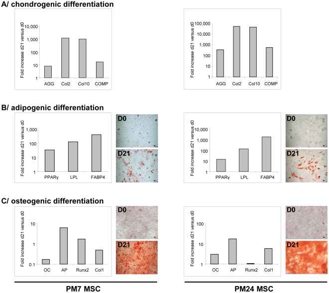
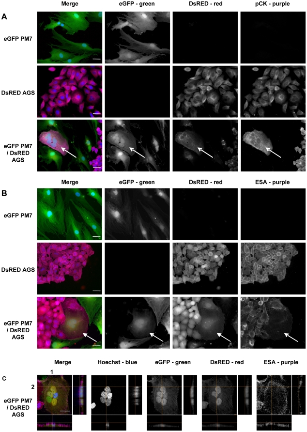
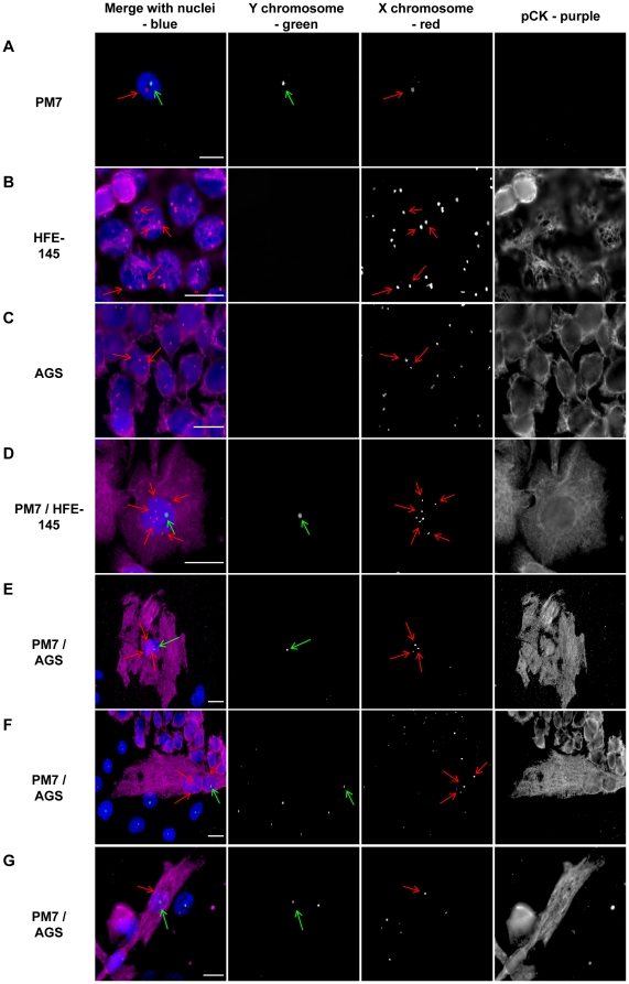
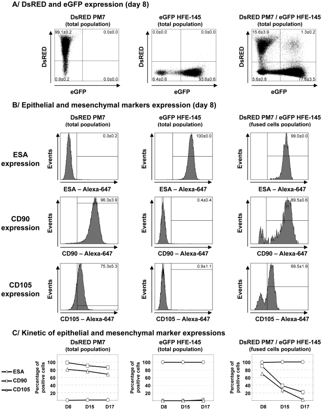
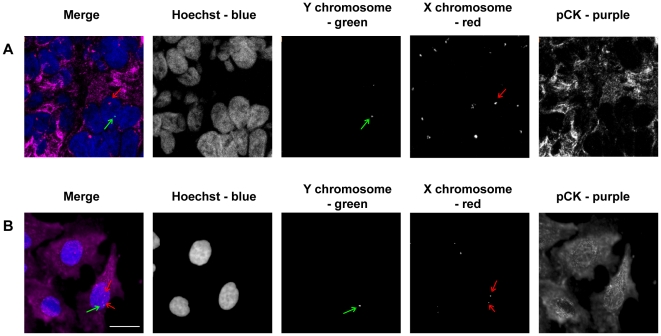
Bone marrow-derived mesenchymal stem cells (MSC) have the ability to differentiate into a variety of cell types and are a potential source for epithelial tissue repair. Several studies have demonstrated their ability to repopulate the gastrointestinal tract (GIT) in bone marrow transplanted patients or in animal models of gastrointestinal carcinogenesis where they were the source of epithelial cancers. However, mechanism of MSC epithelial differentiation still remains unclear and controversial with trans-differentiation or fusion events being evoked. This study aimed to investigate the ability of MSC to acquire epithelial characteristics in the particular context of the gastrointestinal epithelium and to evaluate the role of cell fusion in this process. In vitro coculture experiments were performed with three gastrointestinal epithelial cell lines and MSC originating from two patients. After an 8 day coculture, MSC expressed epithelial markers. Use of a semi-permeable insert did not reproduce this effect, suggesting importance of cell contacts. Tagged cells coculture or FISH on gender-mismatched cells revealed clearly that epithelial differentiation resulted from cellular fusion events, while expression of mesenchymal markers on fused cells decreased over time. In vivo cell xenograft in immunodeficient mice confirmed fusion of MSC with gastrointestinal epithelial cells and self-renewal abilities of these fused cells. In conclusion, our results indicate that fusion could be the predominant mechanism by which human MSC may acquire epithelial characteristics when in close contact with epithelial cells from gastrointestinal origin . These results could contribute to a better understanding of the cellular and molecular mechanisms allowing MSC engraftment into the GIT epithelium.


Conflict of interest statement

Competing Interests: The authors have declared that no competing interests exist.

Figures

Figure 1
Figure 1. Trilineage differentiation of MSC.
Left panels show differentiation of PM7 and right panels show that of PM24. A/ Chondrogenic differentiation was visualized by detection of AGG, Col2, Col10 and COMP mRNAs using RT-PCR. B/ Adipogenic differentiation was visualized by PPAR γ, LPL and FABP4 mRNAs using RT-PCR and Oil Red O staining of lipid droplets on day 0 (D0) and 21 (D21). C/ Osteogenic differentiation was visualized by OC, AP, Runx2 and Col1 mRNAs using RT-PCR and Alizarin Red S staining on day 0 (D0) and 21 (D21). RT-PCR were performed on day 21 and results were compared to undifferentiated MSC on day 0 and normalized with RSP9 mRNAs. One representative experiment out of three is presented. Original magnification ×50. Scale bar, 10 µm.
Figure 2
Figure 2. Immunofluorescent staining of epithelial markers in MSC cocultured with gastric epithelial cells in vitro.
eGFP MSC (PM7) cells were cocultured with DsRED AGS epithelial cells for 8 days. A/ Immunostaining with cytokeratins (pCK) or B/ ESA primary antibodies were revealed with AlexaFluor 647 labelled secondary antibodies (purple), and nuclei were stained with Hoechst 33342 compound (blue). C/ Three-dimensional reconstruction of confocal laser microscopy imaging. Images shown represent maximum intensity projection on the x–y axis of the z-stack and the projections of the orthogonal sections (1 and 2 dotted white lines) of the z-stack at the right side of each image. The first vertical panel shows colored merge images with Hoechst, whereas black and white channels alone follow. White arrows show MSC fused with epithelial cells and expressing cytokeratins. One representative experiment out of three is presented. Scale bar, 10 µm.
Figure 3
Figure 3. FISH analysis of MSC and epithelial gastric cell line cocultures.
eGFP male MSC (PM7) cells were cultured with DsRED female epithelial AGS or HFE-145 cells for 8 days and fixed. FISH (SpectrumGreen-Y chromosome and SpectrumOrange-X chromosome) was performed and pancytokeratin primary antibodies were revealed with AlexaFluor 647 labelled secondary antibodies (purple), and nuclei were stained with Hoechst 33342 compound (blue). The first vertical panel shows colored merge images with Hoechst, whereas black and white channels alone follow. Green arrows show Y chromosomes and red arrows X chromosomes. A–C/ Male PM7 expressed one Y chromosome and one X chromosome whereas female AGS or HFE-145 cells expressed only X chromosomes and cytokeratins. D–E/ In PM7/HFE-145 or AGS cocultured cells: example of one cell expressing cytokeratins and possessing a Y chromosome corresponding to a MSC-derived cell. In PM7/AGS cocultured cells, examples of one cell F/ expressing cytokeratins and harboring one male and one female nuclei or G/ expressing cytokeratins and harboring one nucleus with one Y chomosome and one X chromosome. One representative experiment out of three is presented. Scale bar, 10 µm.
Figure 4
Figure 4. Quantification of fused MSC by flow cytometry.
DsRED MSC (PM7) cells were cultured with eGFP epithelial HFE-145 cells for indicated times, harvested, stained with anti-ESA antibodies, detected by Alexa-647 labelled secondary antibodies and analyzed by flow cytometry. A/ DsRED and eGFP expression of PM7, HFE-145 and cocultured cells was analyzed at day 9. Coculture of DsRED PM7 and eGFP HFE-145 cells led to the appearance of fused cells (1.3%±0.2 of total cells). B/ Fused cells expression of epithelial (ESA) and mesenchymal (CD90 and CD105) markers was measured at day 9. Fused cells expressed ESA (99%±0.0 of positive cells); CD90 (89.5%±0.6) and CD105 (69.5%±1.9). C/ Expression of epithelial and mesenchymal markers was assessed at day 9, 15 and 17. Expression of ESA was stable whereas expression of mesenchymal markers decreased over time. Results represent the mean ± SD of one experiment with three replicates representative of at least three different experiments.
Figure 5
Figure 5. FISH analysis of chromosome content of tumor cells resulting from human MSC and AGS cell xenografts in immunodeficient mice.
A/ Male MSC (PM7) and female AGS cells were injected subcutaneously in NOG mice. After 50 days, mice were sacrificed and tumors resulting from transplanted cells were processed and analyzed by immunofluorescence and FISH (SpectrumGreen-Y chromosome and SpectrumOrange-X chromosome). Cytokeratins were stained with primary antibodies revealed by secondary AlexaFluor 647 labelled antibodies (purple), and nuclei were stained with Hoechst 33342 compound (blue). The first vertical panel shows colored merge images, whereas black and white channels alone follow. Red and green arrows show chromosomes of one male-derived cell expressing cytokeratins. B/ Cells from tumors were dissociated and cultured in vitro for 24 h. Cells were processed as in A. Male derived cells expressing cytokeratins possess abnormal chromosome content but are viable and adhere to coverslips. White bar, 10 µm.

References

    1. Okamoto R, Watanabe M. Molecular and clinical basis for the regeneration of human gastrointestinal epithelia. J Gastroenterol. 2004;39:1–6. - PubMed
    1. Hutchinson L, Stenstrom B, Chen D, Piperdi B, Levey S, et al. Human Barrett's Adenocarcinoma of the Esophagus, Associated Myofibroblasts, and Endothelium Can Arise from Bone Marrow-Derived Cells After Allogeneic Stem Cell Transplant. Stem Cells Dev. 2010;20:11–17. - PMC - PubMed
    1. Korbling M, Katz RL, Khanna A, Ruifrok AC, Rondon G, et al. Hepatocytes and epithelial cells of donor origin in recipients of peripheral-blood stem cells. N Engl J Med. 2002;346:738–746. - PubMed
    1. Matsumoto T, Okamoto R, Yajima T, Mori T, Okamoto S, et al. Increase of bone marrow-derived secretory lineage epithelial cells during regeneration in the human intestine. Gastroenterology. 2005;128:1851–1867. - PubMed
    1. Okamoto R, Yajima T, Yamazaki M, Kanai T, Mukai M, et al. Damaged epithelia regenerated by bone marrow-derived cells in the human gastrointestinal tract. Nat Med. 2002;8:1011–1017. - PubMed

Publication types

MeSH terms