

Overexpression of prothymosin alpha predicts poor disease outcome in head and neck cancer
- PMID: 21573209
- PMCID: PMC3088661
- DOI: 10.1371/journal.pone.0019213
Overexpression of prothymosin alpha predicts poor disease outcome in head and neck cancer
Abstract
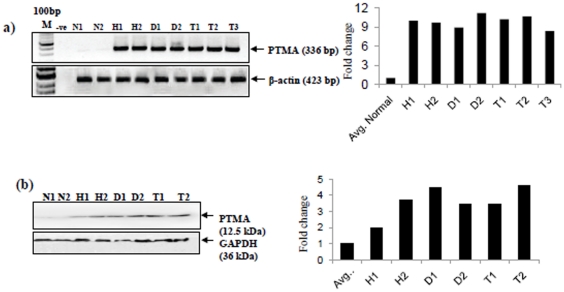
Background: In our recent study, tissue proteomic analysis of oral pre-malignant lesions (OPLs) and normal oral mucosa led to the identification of a panel of biomarkers, including prothymosin alpha (PTMA), to distinguish OPLs from histologically normal oral tissues. This study aimed to determine the clinical significance of PTMA overexpression in oral squamous cell hyperplasia, dysplasia and head and neck squamous cell carcinoma (HNSCC).
Methodology: Immunohistochemistry of PTMA protein was performed in HNSCCs (n = 100), squamous cell hyperplasia (n = 116), dysplasia (n = 50) and histologically normal oral tissues (n = 100). Statistical analysis was carried out to determine the association of PTMA overexpression with clinicopathological parameters and disease prognosis over 7 years for HNSCC patients.
Results: Our immunohistochemical analysis demonstrated significant overexpression of nuclear PTMA in squamous cell hyperplasia (63.8%), dysplasia (50%) and HNSCC (61%) in comparison with oral normal mucosa (p(trend)<0.001). Chi-square analysis showed significant association of nuclear PTMA with advanced tumor stages (III+IV). Kaplan Meier survival analysis indicated reduced disease free survival (DFS) in HNSCC patients (p<0.001; median survival 11 months). Notably, Cox-multivariate analysis revealed nuclear PTMA as an independent predictor of poor prognosis of HNSCC patients (p<0.001, Hazard's ratio, HR = 5.2, 95% CI = 2.3-11.8) in comparison with the histological grade, T-stage, nodal status and tumor stage.
Conclusions: Nuclear PTMA may serve as prognostic marker in HNSCC to determine the subset of patients that are likely to show recurrence of the disease.
Conflict of interest statement
Figures


Similar articles
-
Nuclear S100A7 is associated with poor prognosis in head and neck cancer.PLoS One. 2010 Aug 3;5(8):e11939. doi: 10.1371/journal.pone.0011939. PLoS One. 2010. PMID: 20689826 Free PMC article.
-
Expression of prothymosin alpha predicts early recurrence and poor prognosis of hepatocellular carcinoma.Hepatobiliary Pancreat Dis Int. 2015 Apr;14(2):171-7. doi: 10.1016/s1499-3872(14)60326-x. Hepatobiliary Pancreat Dis Int. 2015. PMID: 25865690
-
Elevated expression of iASPP in head and neck squamous cell carcinoma and its clinical significance.Med Oncol. 2012 Dec;29(5):3381-8. doi: 10.1007/s12032-012-0306-9. Epub 2012 Jul 20. Med Oncol. 2012. PMID: 22815155
-
Increased expression of prothymosin-α, independently or combined with TP53, correlates with poor prognosis in colorectal cancer.Int J Clin Exp Pathol. 2014 Jul 15;7(8):4867-76. eCollection 2014. Int J Clin Exp Pathol. 2014. PMID: 25197357 Free PMC article.
-
Prothymosin alpha expression in the vertebrate testis: a comparative review.Zygote. 2017 Dec;25(6):760-770. doi: 10.1017/S096719941700065X. Epub 2017 Nov 27. Zygote. 2017. PMID: 29173229 Review.
Cited by
-
Expression of prothymosin α in lung cancer is associated with squamous cell carcinoma and smoking.Oncol Lett. 2019 Jun;17(6):5740-5746. doi: 10.3892/ol.2019.10248. Epub 2019 Apr 12. Oncol Lett. 2019. PMID: 31105795 Free PMC article.
-
EZH2 knockdown in tamoxifen-resistant MCF-7 cells unravels novel targets for regaining sensitivity towards tamoxifen.Breast Cancer. 2021 Mar;28(2):355-367. doi: 10.1007/s12282-020-01166-0. Epub 2020 Sep 29. Breast Cancer. 2021. PMID: 32990923
-
Identification of Potential Biomarkers Using Integrative Approach: A Case Study of ESCC.SN Comput Sci. 2023;4(2):114. doi: 10.1007/s42979-022-01492-4. Epub 2022 Dec 21. SN Comput Sci. 2023. PMID: 36573207 Free PMC article.
-
Subcellular dissemination of prothymosin alpha at normal physiology: immunohistochemical vis-a-vis western blotting perspective.BMC Physiol. 2016 Mar 1;16:2. doi: 10.1186/s12899-016-0021-4. BMC Physiol. 2016. PMID: 26932824 Free PMC article.
-
First Evidence of the Expression and Localization of Prothymosin α in Human Testis and Its Involvement in Testicular Cancers.Biomolecules. 2022 Aug 31;12(9):1210. doi: 10.3390/biom12091210. Biomolecules. 2022. PMID: 36139050 Free PMC article.
References
-
- Dosil M, Alvarez-Fernandez L, Gomez-Marquez J. Differentiation-linked expression of prothymosin alpha gene in human myeloid leukemic cells. Exp Cell Res. 1993;204:94–101. - PubMed
-
- Jiang X, Kim HE, Shu H, Zhao Y, Zhang H, et al. Distinctive roles of PHAP proteins and prothymosin-alpha in a death regulatory pathway. Science. 2003;299:223–226. - PubMed
Publication types
MeSH terms
Substances
Grants and funding
LinkOut - more resources
Full Text Sources
Other Literature Sources
Medical
