. 2011 May 5;6(5):e19437.
doi: 10.1371/journal.pone.0019437.

Electromagnetic field effect or simply stress? Effects of UMTS exposure on hippocampal longterm plasticity in the context of procedure related hormone release

Affiliations

Electromagnetic field effect or simply stress? Effects of UMTS exposure on hippocampal longterm plasticity in the context of procedure related hormone release

Nora Prochnow et al. PLoS One. .

Abstract

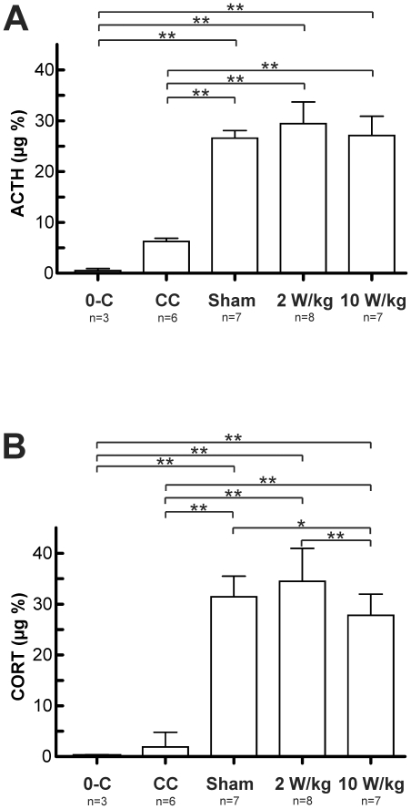
Harmful effects of electromagnetic fields (EMF) on cognitive and behavioural features of humans and rodents have been controversially discussed and raised persistent concern about adverse effects of EMF on general brain functions. In the present study we applied radio-frequency (RF) signals of the Universal Mobile Telecommunications System (UMTS) to full brain exposed male Wistar rats in order to elaborate putative influences on stress hormone release (corticosteron; CORT and adrenocorticotropic hormone; ACTH) and on hippocampal derived synaptic long-term plasticity (LTP) and depression (LTD) as electrophysiological hallmarks for memory storage and memory consolidation. Exposure was computer controlled providing blind conditions. Nominal brain-averaged specific absorption rates (SAR) as a measure of applied mass-related dissipated RF power were 0, 2, and 10 W/kg over a period of 120 min. Comparison of cage exposed animals revealed, regardless of EMF exposure, significantly increased CORT and ACTH levels which corresponded with generally decreased field potential slopes and amplitudes in hippocampal LTP and LTD. Animals following SAR exposure of 2 W/kg (averaged over the whole brain of 2.3 g tissue mass) did not differ from the sham-exposed group in LTP and LTD experiments. In contrast, a significant reduction in LTP and LTD was observed at the high power rate of SAR (10 W/kg). The results demonstrate that a rate of 2 W/kg displays no adverse impact on LTP and LTD, while 10 W/kg leads to significant effects on the electrophysiological parameters, which can be clearly distinguished from the stress derived background. Our findings suggest that UMTS exposure with SAR in the range of 2 W/kg is not harmful to critical markers for memory storage and memory consolidation, however, an influence of UMTS at high energy absorption rates (10 W/kg) cannot be excluded.


Conflict of interest statement

Competing Interests: The authors have declared that no competing interests exist.

Figures

Figure 1
Figure 1. Technical implementations of full brain UMTS exposure.
Figures 1A–C represent the technical implementation of the full-brain UMTS exposure device for six 12–15 weeks aged male Wistar rats. 1A depicts the complete exposure device with 1) the feed, 2) a 1:6 power splitter, 3) the hexagonal double-cone waveguide, 4) 6 shielded exposure compartments, 5) 6 restrainers with holders, 6) the ground plate. Prior to electromagnetic field exposure, animals were inserted into cone-shaped body restrainers to avoid unwanted head and/or body movements (1B, upper image) and subsequently placed and fixed under the EMF exposure cone (1B, lower image). Note that the brain of the animal within the restrainer is located below the lower end of the pair of metal ribs built into each compartment of the double-cone waveguide. 1C is a photograph of the rat cadaver in the restrainer with the fibre-optic temperature probe contacted to the brain tissue through a bore in the cranial bone. Fig. 1D represents the recording and stimulus electrode position for excitatory field potential (fEPSP) recordings of CA1 pyramidal cells: Electrical stimulation (Stim) was applied to the Schaffer collateral pathway to elicit fEPSPs on the dendritic site of CA1 neurons. LTP was evoked by using high frequency stimulation (HFS) with four trains of 10 shocks at 100 Hz every 1 sec. Low frequency stimulation (LFS) for LTD recording was set to one trail of 900 shocks at 1 Hz.
Figure 2
Figure 2. Results of the numerical and experimental dosimetry concerning the RF exposure.
Figure 2A–C shows results of the numerical and experimental dosimetry in order to characterize the RF exposure. Fig. 2A gives computed results of the electrical field (2A, left image) and SAR distributions (2A, right image) indicating the local character of the rat's RF exposure. In Figs. 2B and 2C the temperature development measured during the pre-study with the fibre-optic temperature probe placed in the brain (Fig. 1C) of rat cadavers and of narcotized animals, respectively, is displayed for 2.3 g-brain-averaged SAR values of 8.2 (left images) and 4.1 W/kg (right images). Each of the curves in Fig. 2B which were achieved with dead animals represents a typical temperature response of 1 individual animal. Long times of investigation with the possibility to observe the reaction of switching the RF field on and off were no problem here due to the use of cadavers. The temperature development shows the expected behavior of passive lossy dielectric bodies with heat convection through its surface as main contribution leading to a steady-state. On the other hand, each of the 6 curves in Figure 2C represents the development of temperature in the brain of an anesthetized individual male, adult Wistar rat after onset of RF exposure. It is assumed that the differing dynamic of the temperature curves is due to different depths of the anesthetical state of the animals.
Figure 3
Figure 3. Measurements of the stress related plasma hormones.
3A–B show the outcome of measurements of the stress related plasma hormones Adrenocorticotrophin (ACTH) and Corticosterone (CORT) in the differentially treated groups of animals. The blood was collected from the trunk after decapitation of the animals. Note, that a handled group (0-C) which was trained to visit the guillotine voluntarily after training was used for reference. 3A, Holm-Sidak comparison of the ACTH levels revealed significantly increased values in cage controls (CC), the SAR 0 W/kg (Sham), 2 W/kg (2 W/kg) and 10 W/kg (10 W/kg) treated animals when compared with the 0-C group. Furthermore all exposure setup treated animals showed strongly increased ACTH levels when compared to the CC group, indicating a high degree of procedure related stress. Comparable findings were made in regarding the plasma CORT levels in 3B. The result clearly underlines the handling and procedure related stress component.
Figure 4
Figure 4. Induced CA1 long term potentiation (LTP) of field potential (fEPSP) slopes.
4A–F LTP following high frequency stimulation (HFS) of the Schaffer collateral -commissural pathway of the Hippocampus in the differentially treated groups of Wistar rats. Reliable LTP of fEPSP slopes and amplitudes (as indicated in the figure insets) was achieved in 20–30 min after the theta burst and maintained for at least 40 min. The grey trace in the insets depicts the fEPSP 30 min post stimulation. In comparison to cage controls (4A) the Sham group (4B) as well as the SAR 2 W/kg exposed group (4C) were matter to pronounced early LTP during the first 20–30 min after HFS, followed by persistent LTP of low amplitude. Similar observations were made for the SAR 10 W/kg treated group (4D): HFS stimulation lead to a 30–40 min lasting phase of early LTP. In this case early LTP could not be shown to be converted into a subsequent phase of late LTP. Scale in insets: horizontal = 5 ms, vertical = 0.5 mV. In 4E, fEPSP slopes between the four groups were compared during the first five minutes of early LTP induction. The process of early LTP is clearly reflected by the significant differences between the differentially treated groups. All exposure setup treated animals differed in their early LTP from the cage control group either in increased fEPSP slopes (Sham, 2 W/kg) or a lack in LTP induction (10 W/kg). Comparison of the late LTP phase >30 min post HFS in 4F reveals significantly reduced fEPSP slopes in 10 W/kg exposed animals compared to all other groups.
Figure 5
Figure 5. Induced CA1 long term depression (LTD) of field potential (fEPSP) slopes.
5A–F LTD following low frequency stimulation (LFS) of the Schaffer collateral -commissural pathway of the Hippocampus in the differentially treated groups of Wistar rats. Sustained LTD of fEPSP slopes was induced in all four groups 20–30 min following LFS and at least maintained for 60 min. The grey trace in the insets depicts the fEPSP 30 min post stimulation. Compared to cage controls (A), Sham and EMF exposure treated animals (B–D) also showed LTD which was characterized by much lower changes in the LFS induced fEPSP responses. The SAR 10 W/kg treated group (D) was characterized by the lowest depression in fEPSP slopes 30 min post LFS. Scale in insets: horizontal = 5 ms, vertical = 0.5 mV. 5E depicts the comparison of fEPSP slopes in the inductive phase of LTD during the first 5 min post LFS. As indicated by the figure insets in 5A–D, all exposure setup treated animals were characterized by significantly flattened fEPSP slopes. This result became strengthened by the comparison of the fEPSP slopes 30 min after LTD induction in 5F: All UMTS exposure setup treated animals exhibited a reduced LTD in which SAR 10 W/kg treated animals differed from the sham group by the weakest induced responses.
Figure 6
Figure 6. Comparison of the input/output function for basal synaptic transmission in slices.
Brains were derived from cage controls and all three exposure setup treated groups. The size of presynaptic fibre volleys (input) was compared to the slopes of fEPSPs (output) measured from responses generated by different intensities (25, 50, 75, 100% of the maximum fEPSP amplitude) of presynaptic fibre stimulation. The 10 W/kg exposed group is characterized by a prominent left shift of their fibre volleys of the fEPSPs corresponding to 75 and 100% of the maximum fEPSP amplitude. Furthermore the group is characterized by general decrease in fEPSP slopes when compared to cage controls. These correlations underline the findings of the general negative influence of the exposure setup treatment on the animals' synaptic transmission properties which might be most probably due to exposure and restraint related stress.
Figure 7
Figure 7. Correlation of stress hormone levels and synaptic plasticity.
7A, levels in plasma corticosterone (CORT) were found to be negatively correlated with the outcome in late LTP of the four differentially treated groups (as indexed in the diagrams). CORT levels were positively correlated with persistent LTD of the groups in 7B. Each data point combines the individual groups' mean CORT and fEPSP slope value for persistent late LTP/LTD.

References

    1. Zwamborn APM, Vossen SHJ, van Leersum BJA, Ouwens MA, Makel WN. Netherlands Organization for Applied Scientific Research (TNO); 2003. Effects of global communication system radio-frequency fields on well being and cognitive functions of human subjects with and without subjective complaints.
    1. Regel SJ, Negovetic S, Roosli M, Berdinas V, Schuderer J, et al. UMTS base station-like exposure, well-being, and cognitive performance. Environ Health Perspect. 2006;114:1270–1275. - PMC - PubMed
    1. Kleinlogel H, Dierks T, Koenig T, Lehmann H, Minder A, et al. Effects of weak mobile phone - electromagnetic fields (GSM, UMTS) on well-being and resting EEG. Bioelectromagnetics. 2008;29:479–487. - PubMed
    1. Unterlechner M, Sauter C, Schmid G, Zeitlhofer J. No effect of an UMTS mobile phone-like electromagnetic field of 1.97 GHz on human attention and reaction time. Bioelectromagnetics. 2008;29:145–153. - PubMed
    1. Eltiti S, Wallace D, Ridgewell A, Zougkou K, Russo R, et al. Short-term exposure to mobile phone base station signals does not affect cognitive functioning or physiological measures in individuals who report sensitivity to electromagnetic fields and controls. Bioelectromagnetics. 2009;30:556–563. - PubMed