. 2011 Oct;45(2):309-20.
doi: 10.1007/s12031-011-9523-5. Epub 2011 May 15.

Protein kinase B (Akt) and mitogen-activated protein kinase p38α in retinal ischemic post-conditioning

Affiliations

Protein kinase B (Akt) and mitogen-activated protein kinase p38α in retinal ischemic post-conditioning

John C Dreixler et al. J Mol Neurosci. 2011 Oct.

Abstract

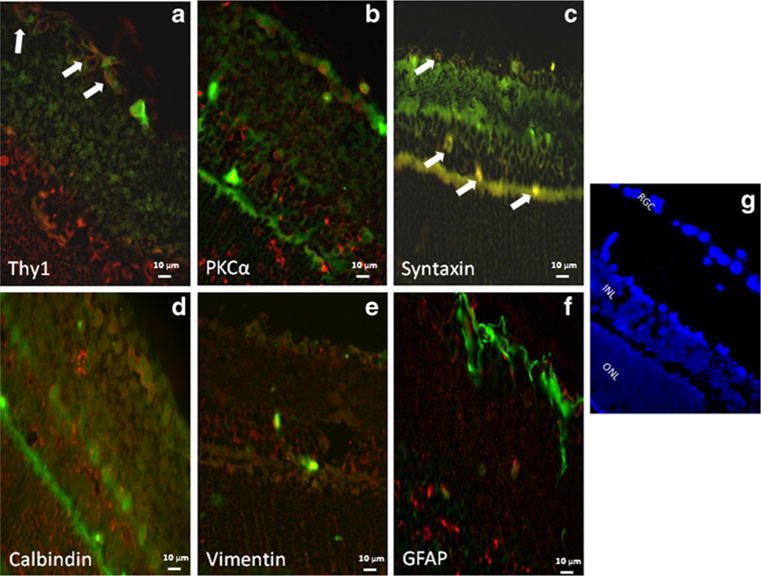
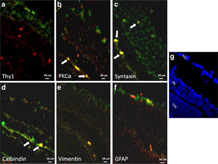
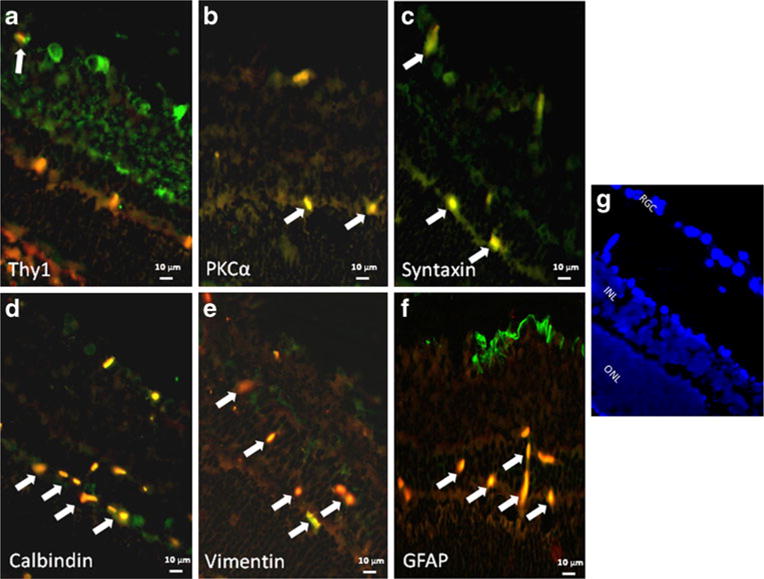
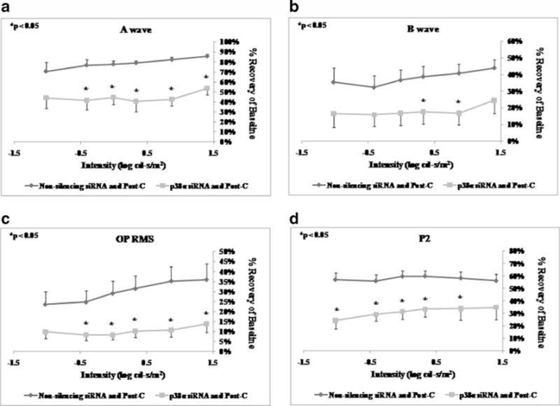
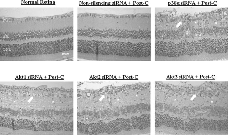
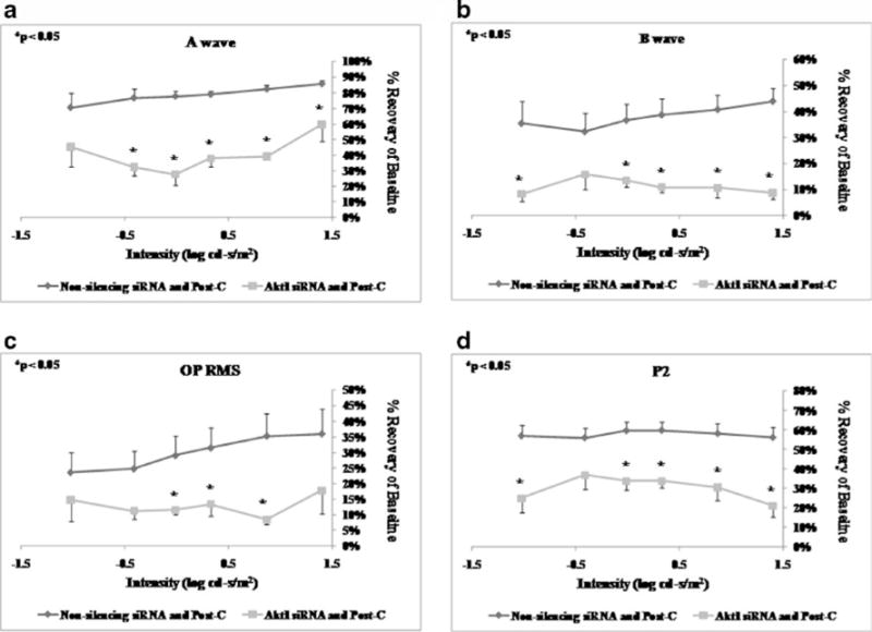
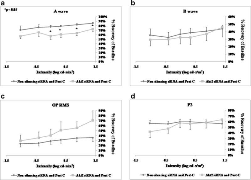
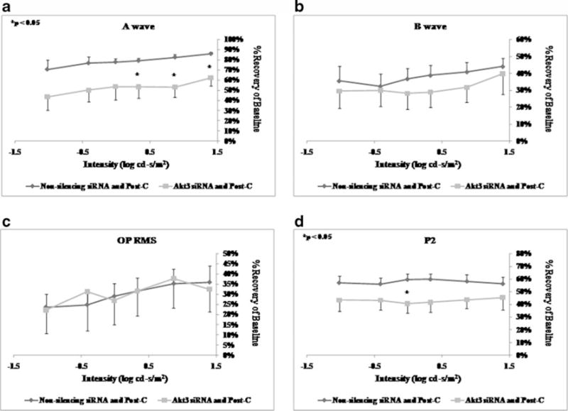
In previous studies, it was shown that post-conditioning, a transient period of brief ischemia following prolonged severe ischemia in the retina, could provide significant improvement in post-ischemic recovery, attenuation of cell loss, and decreased apoptosis. However, the mechanisms of post-conditioning in the retina have not been elucidated. We hypothesized that two kinases, mitogen-activated protein kinase p38α and protein kinase B (Akt), were involved in the mechanism of post-conditioning. Ischemia was induced in rat retina in vivo. Recovery after ischemia followed by 8 min of post-conditioning early in the reperfusion period after prolonged ischemia was assessed functionally (electroretinography) and histologically at 7 days after ischemia. We examined the role of p38α and Akt subtypes 1-3 in post-conditioning by intravitreal injection of interfering RNA 6 h prior to ischemia and post-conditioning and compared the results to injection of non-silencing interfering RNA sequence. The blockade of p38α significantly decreased the recovery after ischemia and post-conditioning, and enhanced cell loss and disorganization of the retina. Blockade of Akt1, and to a lesser degree, Akt2, significantly decreased the recovery after ischemia and enhanced cell loss and disorganization. These differences in the effects of blockade of Akt subtypes were not explainable by distribution of Akt subtypes in the retina, which were similar. In conclusion, both p38 and Akt are essential components of the neuroprotection induced by post-ischemic conditioning in the retina.


Conflict of interest statement

There is no conflict of interest or commercial interest for any of the authors.

Figures

Fig. 1
Fig. 1
Co-localization of Akt1 (red) and retinal cell markers (green). Co-localization (as depicted by an orange/yellow color) of Akt1with the retinal ganglion cell marker, anti-Thy1 (a) and with amacrine cell marker, anti-syntaxin (c) is indicated by white arrows. The fluorescent images utilized ×40 oil magnification. Control sections for primary antibodies incubated with non-immune serum demonstrated no staining (not shown). Scale bars are shown at the bottom of each figure. Orientation of the retinal layers are shown in (g) by the nuclear staining by DAPI. The figure shows that Akt1 is present primarily in cells in the retinal ganglion cell layer
Fig. 2
Fig. 2
Co-localization of Akt2 (red) and retinal cell markers (green). Co-localization (as depicted by an orange/yellow color) of Akt2 with bipolar cell marker, anti-PKCα (b), with amacrine cell marker, anti- syntaxin (c), and with horizontal cell marker, anti-calbindin (d), is indicated by white arrows. The fluorescent images utilized ×40 oil magnification. Control sections for primary antibodies incubated with non-immune serum demonstrated no staining (not shown). Scale bars are shown at the bottom of each figure. Orientation of the retinal layers are shown in (g) by the nuclear staining by DAPI. The figure shows that Akt2 is present in amacrine cells (including displaced amacrine cells in the RGC layer), bipolar cells, and horizontal cells. We also note that some apparent Akt2 co-localization with astrocytic markers may be attributed to non-specific staining
Fig. 3
Fig. 3
Co-localization of Akt3 (red) and retinal cell markers (green). Co-localization (as depicted by an orange/yellow color) of Akt3 with the retinal ganglion cell marker, anti-Thy1 (a), with bipolar cell marker, anti-PKCα (b), with amacrine cell marker, anti-syntaxin (c), with horizontal cell marker, anti-calbindin (d), and with astrocyte/ Müller cell marker, anti-vimentin (e) and anti-GFAP (f), is indicated by white arrows. The fluorescent images utilized ×40 oil magnification. Control sections for primary antibodies incubated with non-immune serum demonstrated no staining (not shown). Scale bars are shown at the bottom of each figure. Orientation of the retinal layers are shown in (g) by the nuclear staining by DAPI. The figure shows that Akt3 is present in nuclei throughout the inner retina and RGCs, amacrine cells, bipolar cells, horizontal cells, and in astrocytes and Müller cells. We also note co-localization of Akt3 with Thy1 and calbindin in unexpected retinal layers that may be attributed to non-specific staining
Fig. 4
Fig. 4
ERG stimulus-intensity response in ischemic post-conditioned retinae treated with siRNA to p38α vs. non-silencing sequence. The recovery relative to baseline at 7 days after ischemia is shown on the y- axis. Double normalized (for baseline in ischemic eye and for variation in the non-ischemic eye) ERG data for a-wave (a), b-wave (b), OP RMS (c), and P2 (d) over a range of flash intensities suggest that p38α is essential in post-conditioning. Solid lines with diamonds=non- silencing siRNA+ischemia+Post-C (n=5). Solid lines with squares= p38α siRNA+ischemia+Post-C (n = 6). Statistical analysis was achieved using unpaired t tests
Fig. 5
Fig. 5
Representative histopathological images of hematoxylin-and- eosin-stained retinae in 4-μm-thick sections for each of the experimental groups. These sections were prepared from retinae removed from the rats at 7 days following ischemia. Arrows indicate layers demonstrating cell loss and asterisks denote regions of inflammatory cell infiltration. The retinal layers and scale bars are depicted in the normal retina image
Fig. 6
Fig. 6
ERG stimulus-intensity response in ischemic post-conditioned retinae treated with siRNA to Akt1 vs. non-silencing sequence. The recovery relative to baseline at 7 days after ischemia is shown on the y-axis. Double normalized (for baseline in ischemic eye and for variation in the non-ischemic eye) ERG data for a-wave (a), b-wave (b), OP RMS (c), and P2 (d) over a range of flash intensities suggest that Akt1 is essential in post-conditioning. Solid lines with diamonds= non-silencing siRNA+ischemia+Post-C (n=5). Solid lines with squares=Akt1 siRNA+ischemia+Post-C (n=6). Statistical analysis was achieved using unpaired t tests
Fig. 7
Fig. 7
ERG stimulus-intensity response in ischemic post-conditioned retinae treated with siRNA to Akt2 vs. non-silencing sequence. The recovery relative to baseline at 7 days after ischemia is shown on the y-axis. Double normalized (for baseline in ischemic eye and for variation in the non-ischemic eye) ERG data for a-wave (a), b-wave (b), OP RMS (c), and P2 (d) over a range of flash intensities suggest that Akt2 has a minor role in post-conditioning. Solid lines with diamonds=non-silencing siRNA+ischemia+Post-C (n=5). Solid lines with squares=Akt2 siRNA+ischemia+Post-C (n=5). Statistical analysis was achieved using unpaired t tests
Fig. 8
Fig. 8
ERG stimulus-intensity response in ischemic post-conditioned retinae treated with siRNA to Akt3 vs. non-silencing sequence. The recovery relative to baseline at 7 days after ischemia is shown on the y-axis. Double normalized (for baseline in ischemic eye and for variation in the non-ischemic eye) ERG data for a-wave (a), b-wave (b), OP RMS (c), and P2 (d) over a range of flash intensities suggest that Akt3 is not involved in post-conditioning. Solid lines with diamonds=non-silencing siRNA+ischemia+Post-C (n = 5). Solid lines with squares=Akt3 siRNA+ischemia+Post-C (n=9). Statistical analysis was achieved using unpaired t tests

References

    1. Bui BV, Edmunds B, Cioffi GA, Fortune B. The gradient of retinal functional changes during acute intraocular pressure elevation. Invest Ophthalmol Vis Sci. 2005;46:202–213. - PubMed
    1. Coulthard LR, White DE, Jones DL, McDermott MF, Burchill SA. p38(MAPK): stress responses from molecular mechanisms to therapeutics. Trends Mol Med. 2009;15:369–379. - PMC - PubMed
    1. Dorsett Y, Tuschl T. siRNAs: applications in functional genomics and potential as therapeutics. Nat Rev. 2004;3:318–329. - PubMed
    1. Dreixler JC, Shaikh AR, Shenoy SK, Shen Y, Roth S. Protein kinase C subtypes and retinal ischemic preconditioning. Exp Eye Res. 2008;87:300–311. - PMC - PubMed
    1. Dreixler JC, Barone FC, Shaikh AR, Du E, Roth S. Mitogen- activated protein kinase p38alpha and retinal ischemic preconditioning. Exp Eye Res. 2009a;89:782–790. - PMC - PubMed

Publication types

MeSH terms

LinkOut - more resources