Correct anteroposterior patterning of the zebrafish neurectoderm in the absence of the early dorsal organizer
- PMID: 21575247
- PMCID: PMC3120780
- DOI: 10.1186/1471-213X-11-26
Correct anteroposterior patterning of the zebrafish neurectoderm in the absence of the early dorsal organizer
Abstract
Background: The embryonic organizer (i.e., Spemann organizer) has a pivotal role in the establishment of the dorsoventral (DV) axis through the coordination of BMP signaling. However, as impaired organizer function also results in anterior and posterior truncations, it is of interest to determine if proper anteroposterior (AP) pattern can be obtained even in the absence of early organizer signaling.
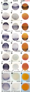
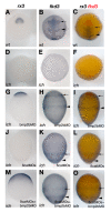
Results: Using the ventralized, maternal effect ichabod (ich) mutant, and by inhibiting BMP signaling in ich embryos, we provide conclusive evidence that AP patterning is independent of the organizer in zebrafish, and is governed by TGFβ, FGF, and Wnt signals emanating from the germ-ring. The expression patterns of neurectodermal markers in embryos with impaired BMP signaling show that the directionality of such signals is oriented along the animal-vegetal axis, which is essentially concordant with the AP axis. In addition, we find that in embryos inhibited in both Wnt and BMP signaling, the AP pattern of such markers is unchanged from that of the normal untreated embryo. These embryos develop radially organized trunk and head tissues, with an outer neurectodermal layer containing diffusely positioned neuronal precursors. Such organization is reflective of the presumed eumetazoan ancestor and might provide clues for the evolution of centralization in the nervous system.
Conclusions: Using a zebrafish mutant deficient in the induction of the embryonic organizer, we demonstrate that the AP patterning of the neuroectoderm during gastrulation is independent of DV patterning. Our results provide further support for Nieuwkoop's "two step model" of embryonic induction. We also show that the zebrafish embryo can form a radial diffuse neural sheath in the absence of both BMP signaling and the early organizer.
Figures


Similar articles
-
Chordin and dickkopf-1b are essential for the formation of head structures through activation of the FGF signaling pathway in zebrafish.Dev Biol. 2017 Apr 15;424(2):189-197. doi: 10.1016/j.ydbio.2017.02.018. Epub 2017 Mar 1. Dev Biol. 2017. PMID: 28259755
-
The homeobox gene bozozok promotes anterior neuroectoderm formation in zebrafish through negative regulation of BMP2/4 and Wnt pathways.Development. 2000 Jun;127(11):2333-45. doi: 10.1242/dev.127.11.2333. Development. 2000. PMID: 10804176
-
The role of the homeodomain protein Bozozok in zebrafish axis formation.Int J Dev Biol. 2001;45(1):299-310. Int J Dev Biol. 2001. PMID: 11291860
-
Dickkopf1 and the Spemann-Mangold head organizer.Int J Dev Biol. 2001;45(1):237-40. Int J Dev Biol. 2001. PMID: 11291852 Review.
-
Specification of anteroposterior axis by combinatorial signaling during Xenopus development.Wiley Interdiscip Rev Dev Biol. 2016 Mar-Apr;5(2):150-68. doi: 10.1002/wdev.217. Epub 2015 Nov 6. Wiley Interdiscip Rev Dev Biol. 2016. PMID: 26544673 Review.
Cited by
-
The history and enduring contributions of planarians to the study of animal regeneration.Wiley Interdiscip Rev Dev Biol. 2013 May-Jun;2(3):301-26. doi: 10.1002/wdev.82. Epub 2012 Jul 23. Wiley Interdiscip Rev Dev Biol. 2013. PMID: 23799578 Free PMC article. Review.
-
Full transcriptome analysis of early dorsoventral patterning in zebrafish.PLoS One. 2013 Jul 29;8(7):e70053. doi: 10.1371/journal.pone.0070053. Print 2013. PLoS One. 2013. PMID: 23922899 Free PMC article.
-
Evolutionarily conserved Wnt/Sp5 signaling is critical for anterior-posterior axis patterning in sea urchin embryos.iScience. 2023 Dec 2;27(1):108616. doi: 10.1016/j.isci.2023.108616. eCollection 2024 Jan 19. iScience. 2023. PMID: 38179064 Free PMC article.
-
Transposon insertion causes ctnnb2 transcript instability that results in the maternal effect zebrafish ichabod (ich) mutation.bioRxiv [Preprint]. 2025 Mar 4:2025.02.28.640854. doi: 10.1101/2025.02.28.640854. bioRxiv. 2025. Update in: Biochim Biophys Acta Gene Regul Mech. 2025 Sep;1868(3):195104. doi: 10.1016/j.bbagrm.2025.195104. PMID: 40093107 Free PMC article. Updated. Preprint.
-
Zebrafish as a model for human epithelial pathology.Lab Anim Res. 2025 Feb 3;41(1):6. doi: 10.1186/s42826-025-00238-6. Lab Anim Res. 2025. PMID: 39901304 Free PMC article. Review.
References
Publication types
MeSH terms
Substances
Grants and funding
LinkOut - more resources
Full Text Sources
Molecular Biology Databases

