. 2011 May 18;100(10):2440-9.
doi: 10.1016/j.bpj.2011.03.062.

Drosophila katanin-60 depolymerizes and severs at microtubule defects

Affiliations

Drosophila katanin-60 depolymerizes and severs at microtubule defects

Juan Daniel Díaz-Valencia et al. Biophys J. .

Abstract

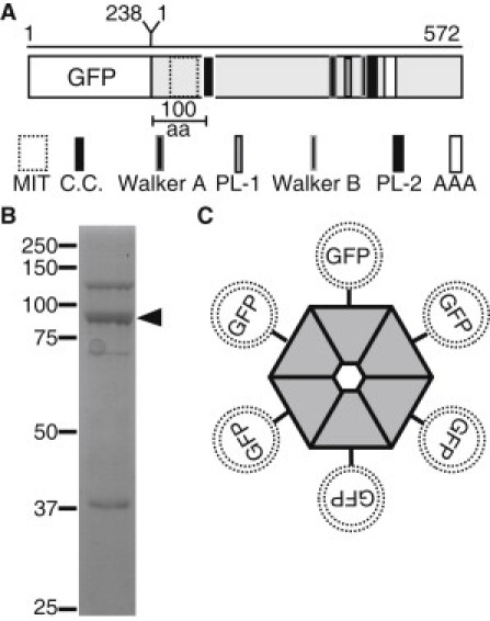
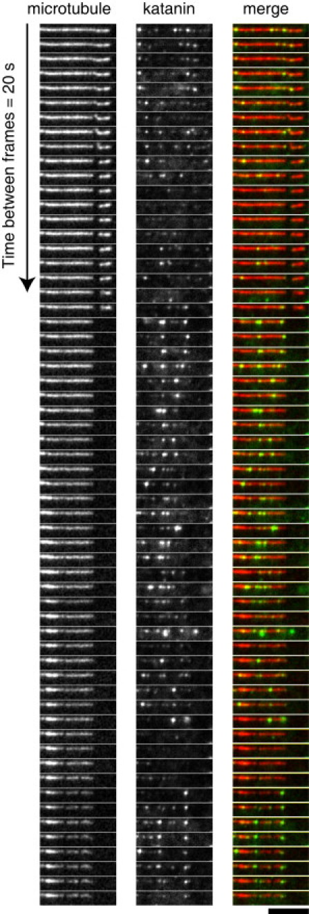
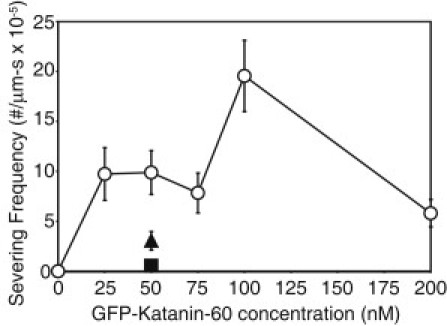
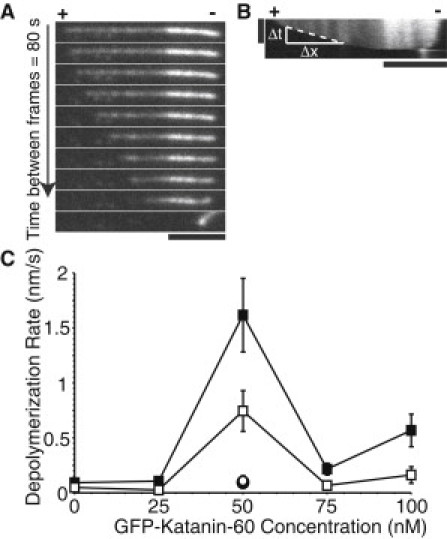
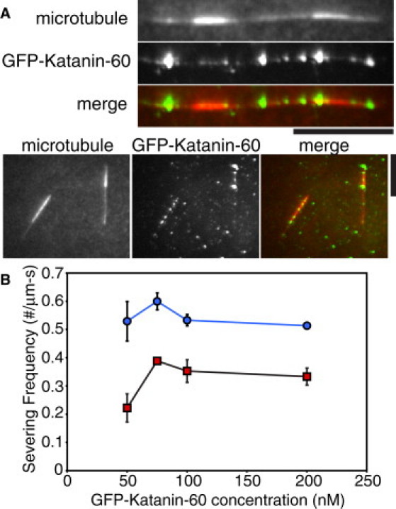
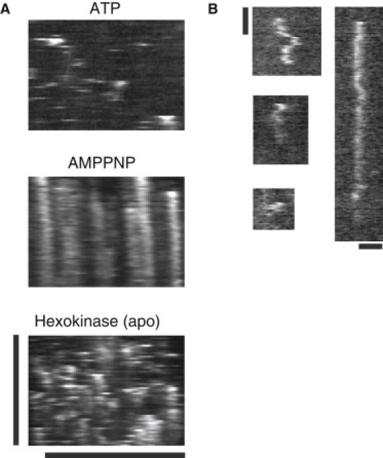
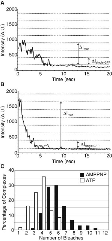
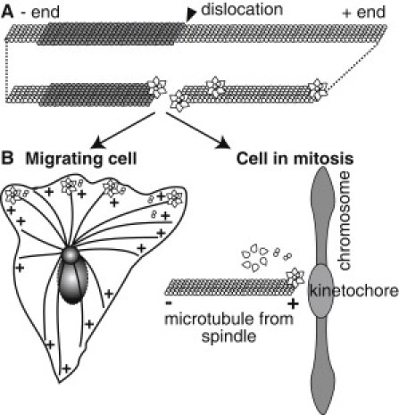
Microtubule (MT) length and location is tightly controlled in cells. One novel family of MT-associated proteins that regulates MT dynamics is the MT-severing enzymes. In this work, we investigate how katanin (p60), believed to be the first discovered severing enzyme, binds and severs MTs via single molecule total internal reflection fluorescence microscopy. We find that severing activity depends on katanin concentration. We also find that katanin can remove tubulin dimers from the ends of MTs, appearing to depolymerize MTs. Strikingly, katanin localizes and severs at the interface of GMPCPP-tubulin and GDP-tubulin suggesting that it targets to protofilament-shift defects. Finally, we observe that binding duration, mobility, and oligomerization are ATP dependent.


Figures

Figure 1
Figure 1
Description of domain architecture and purification of GFP-Katanin-60 from Sf9 insect cells. (A) Schematic depiction, approximately to scale, of domain architecture of GFP-Katanin-60. The amino acid length of GFP and Katanin-60 are indicated at the top of the figure. (aa, amino acids; MIT, microtubule interaction and trafficking domain; C.C., coiled-coil; PL-1, Pore Loop-1; PL-2, Pore Loop-2; AAA, AAA minimum consensus ATPase domain, Walker A, Walker B) (B) A coomassie stained SDS-PAGE gel of purified 6×His-tagged GFP-Katanin-60 after protein purification. Arrow marks the position of GFP-Katanin-60 (92 kD) that is the major band and molecular mass markers in kDa indicated at the left. (C) Cartoon of hexameric GFP-Katanin-60 ring showing the location of GFP at NH-terminal.
Figure 2
Figure 2
Localization of GFP-Katanin-60 during activity on MTs. Time series of MT-severing assays using rhodamine polarity-marked MTs with 200 nM GFP-Katanin-60 at 20 s intervals. From left to right, MTs (red in merge), GFP-Katanin-60 (green in merge), and merge. Scale bar, 5 μm.
Figure 3
Figure 3
Quantitative measurement of GFP-Katanin-60 MT severing expressed as severing frequency of MTs in the presence of 2 mM ATP [0 nM (N = 18); 25 nM (N = 39); 50 nM (N = 80); 75 nM (N = 48); 100 nM (N = 34); 200 nM (N = 77), circles], in the presence of AMPPNP (N = 11, triangle) in the presence of hexokinase to use up residual ATP (N = 10, square). N-values represent the number of MTs analyzed, the points on the plot represent the mean value, and the error bars represent the mean ± SE.
Figure 4
Figure 4
GFP-Katanin-60 depolymerizes MTs in an ATP and concentration dependent manner. (A) Time series of rhodamine polarity-marked MTs with 50 nM GFP-Katanin-60. The (+) indicates the plus end and the (−) indicates the minus end. Time between frames is 80 s; scale bar, 5 μm. (B) Kymograph from MT showing depolymerization is faster at plus end (+). Kymographs were used to measure depolymerization rates by drawing a line and measuring the change in distance over the change in time. Vertical scale bar, 10 min; horizontal scale bar, 5 μm. (C) Quantitative measurement of GFP-Katanin-60 MT depolymerization expressed as rate of depolymerization of MTs at plus end [0 nM (N = 19), 25 nM (N = 39), 50 nM (N = 77), 75 nM (N = 53), 100 nM (N = 76), black squares], minus end [0 nM (N = 19), 25 nM (N = 39), 50 nM (N = 164), 75 nM (N = 49), 100 nM (N = 66), white squares] in the presence of 2 mM ATP, with AMPPNP at plus end (N = 12, white circle), and hexokinase at plus end (N = 16, black circle). N-values represent the number of MTs analyzed, the points on the plot represent the mean value, and the error bars represent the mean ± SE.
Figure 5
Figure 5
GFP-Katanin-p60 severs at interfaces between GMPCPP and GDP-taxol MT segments. (A) The MT frames depict the MTs as they existed at the beginning of the movie before severing (red in merge). The GFP-Katanin-60 frame is a z-projection of the SD of a time series of GFP-Katanin-60 binding (green in merge). Scale bars, 5 μm. (B) Measurement of frequency of severing at 50 nM (N = 9), 75 nM (N = 18), 100 nM (N = 17), and 200 nM (N = 21) GFP-Katanin-60 at interfaces (blue circles) and 1 μm away from interfaces (red squares). N-values represent the number of MTs analyzed, the points on the plot represent the mean value, and the error bars represent the mean ± SE.
Figure 6
Figure 6
GFP-Katanin-60 dynamics depends on the nucleotide state. (A) Kymographs of 50 nM GFP-Katanin-60 in the presence of 2 mM ATP, 5 mM AMPPNP, and 5 mM hexokinase. Vertical scale bar, 20 min; horizontal scale bar, 5 μm for all images. (B) Examples of kymographs of GFP-Katanin-60 diffusing along taxol-stabilized MTs in the presence of 2 mM ATP. Vertical scale bar, 1 min; horizontal scale bar, 1 μm.
Figure 7
Figure 7
Photobleaching and fluorescence intensity analysis of GFP-Katanin-60. (A) Representative example of three-step photobleaching event in the presence of 2 mM ATP. (B) Example of multiple photobleaching events in the presence of 2 mM AMPPNP. (C) Probability distribution of number of bleach events per complex in the presence of 2 mM ATP (white bars) and 2 mM AMPPNP (black bars). The average number of bleaching events is 4/complex for ATP; whereas the average number of bleaching events is 6/complex for AMPPNP.
Figure 8
Figure 8
Illustration of proposed GFP-Katanin-60 activities in vitro and in the cellular context. (A) The GFP-Katanin-60 assembles as a hexamer in the presence of ATP, and associates with defects in the lattice, such as protofilament shifts and MT ends. Once GFP-Katanin-60 binds, it rapidly removes dimers from active sites at plus ends and at defects. (B) In the cell, the same katanin could regulate MT plus ends to alter length and dynamics at the leading edge of migrating cells or at the kinetochores in the mitotic spindle.

References

    1. Conde C., Cáceres A. Microtubule assembly, organization and dynamics in axons and dendrites. Nat. Rev. Neurosci. 2009;10:319–332. - PubMed
    1. Desai A., Mitchison T.J. Microtubule polymerization dynamics. Annu. Rev. Cell Dev. Biol. 1997;13:83–117. - PubMed
    1. Valiron O., Caudron N., Job D. Microtubule dynamics. Cell. Mol. Life Sci. 2001;58:2069–2084. - PMC - PubMed
    1. Roll-Mecak A., McNally F.J. Microtubule-severing enzymes. Curr. Opin. Cell Biol. 2010;22:96–103. - PMC - PubMed
    1. Hartman J.J., Mahr J., McNally F.J. Katanin, a microtubule-severing protein, is a novel AAA ATPase that targets to the centrosome using a WD40-containing subunit. Cell. 1998;93:277–287. - PubMed

Publication types

MeSH terms

LinkOut - more resources