. 2011 May 18;100(10):2450-6.
doi: 10.1016/j.bpj.2011.04.015.

Kinesin walks the line: single motors observed by atomic force microscopy

Affiliations

Kinesin walks the line: single motors observed by atomic force microscopy

Iwan A T Schaap et al. Biophys J. .

Abstract

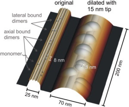
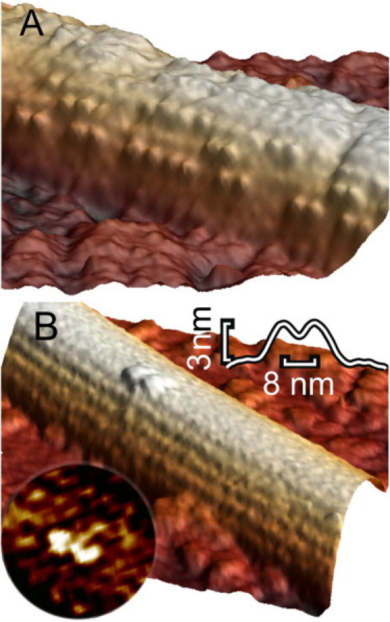
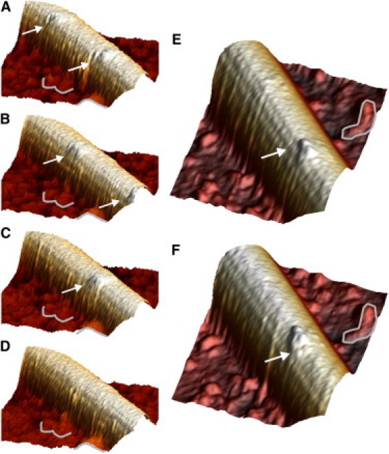
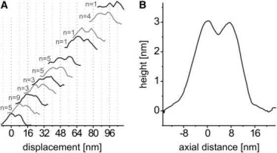
Motor proteins of the kinesin family move actively along microtubules to transport cargo within cells. How exactly a single motor proceeds on the 13 narrow lanes or protofilaments of a microtubule has not been visualized directly, and there persists controversy on the relative position of the two kinesin heads in different nucleotide states. We have succeeded in imaging Kinesin-1 dimers immobilized on microtubules with single-head resolution by atomic force microscopy. Moreover, we could catch glimpses of single Kinesin-1 dimers in their motion along microtubules with nanometer resolution. We find in our experiments that frequently both heads of one dimer are microtubule-bound at submicromolar ATP concentrations. Furthermore, we could unambiguously resolve that both heads bind to the same protofilament, instead of straddling two, and remain on this track during processive movement.


Figures

Figure 1
Figure 1
Simulation of the tip-sample dilation occurring in imaging a MT decorated with kinesin. The left image shows a MT with 25 nm diameter and 13 protofilaments. The MT is decorated with kinesin motors in different orientations, with either both heads bound to two parallel protofilaments, along one protofilament, or with just one single head bound. The right image shows the dilation caused by scanning with a 15 nm parabolic tip. The lateral dimensions get exaggerated by the tip-sample dilation, but the heads remain clearly visible and their axial spacing is not affected.
Figure 2
Figure 2
Individual kinesin motors immobilized on MTs in the presence of 15 μM AMP-PNP. Both images are 3D-rendered. (A) Intermediate kinesin concentration (<1 motor/tubulin dimer). Individual motors could be clearly distinguished. Heads always appeared in pairs, aligned parallel to the MT axis. (B) Low kinesin concentration (<1 motor/10 tubulin dimers). Isolated motors could be seen. Both heads were bound to the same protofilament. (Upper inset) Averaged axial profile of 17 kinesin molecules, interhead spacing: 8 nm, height: 3.0 nm. (Lower inset) Laplacian filtered image of another kinesin molecule showing clearly the two heads and a ∼10 nm long structure that might represent the stalk.
Figure 3
Figure 3
Single kinesin motors moving along a MT. (Movies S1 and S2). (AD) Four frames (30 s acquisition time per frame) of a movie: This was the only movie in which we observed two kinesin motors moving; see Fig S2 for a top-view rendering. The average speed was 3 nm/s at 0.5 μM ATP. (A) Two kinesin dimers, indicated by arrows. (B) Both motors have proceeded (only the last head is still visible of the leading dimer). (C) The second kinesin has proceeded further. (D) The second kinesin has also disappeared. (E and F) Two frames from another sample show a displacement of 16 nm. The two kinesin heads are clearly visible. Fiduciary marks on the background were used to correct for drift. The position of the arrow is fixed in both frames with respect to the fiduciary marks.
Figure 4
Figure 4
Aligned topographical profiles (parallel to the protofilament axis) of moving kinesin dimers, showing the 8 nm periodicity of the resting positions. (A) Displacements found for the 20 isolated moving kinesin dimers, recorded with different samples. Each event comprised 2 to 4 frames. Each curve represents a profile taken along the protofilament with kinesin bound. The position of the profiles from a single movie was referenced to fiduciary marks in the background to correct for drift. The x axis shows the axial displacement of the respective kinesin compared to the first or any of the previous frames. Most displacement distances were observed more than once, in which case the curves were averaged. Curves are plotted with a vertical offset for clarity. (B) Average of all profiles after subtracting the respective multiple of 8 nm. The heads, both bound to the MT and spaced 8 nm apart, can be clearly distinguished.

References

    1. Alberts B., Johnson A., Walter P. Garland Science; New York: 2008. Molecular Biology of the Cell.
    1. Vale R.D., Milligan R.A. The way things move: looking under the hood of molecular motor proteins. Science. 2000;288:88–95. - PubMed
    1. Veigel C., Schmidt C.F. Moving into the cell: single-molecule studies of molecular motors in complex environments. Nat. Rev. Mol. Cell Biol. 2011;12:163–176. - PubMed
    1. Kuo S.C., Gelles J., Sheetz M.P. A model for kinesin movement from nanometer-level movements of kinesin and cytoplasmic dynein and force measurements. J. Cell Sci. Suppl. 1991;14:135–138. - PubMed
    1. Ray S., Meyhöfer E., Howard J. Kinesin follows the microtubule's protofilament axis. J. Cell Biol. 1993;121:1083–1093. - PMC - PubMed

Publication types

MeSH terms

LinkOut - more resources