. 2011 May 19;9(5):363-75.
doi: 10.1016/j.chom.2011.04.008.

IFI16 acts as a nuclear pathogen sensor to induce the inflammasome in response to Kaposi Sarcoma-associated herpesvirus infection

Affiliations

IFI16 acts as a nuclear pathogen sensor to induce the inflammasome in response to Kaposi Sarcoma-associated herpesvirus infection

Nagaraj Kerur et al. Cell Host Microbe. .

Abstract

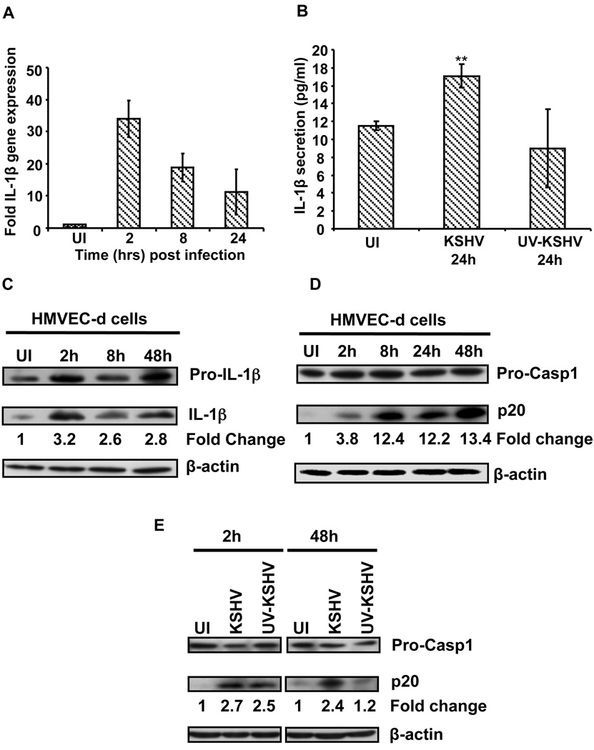
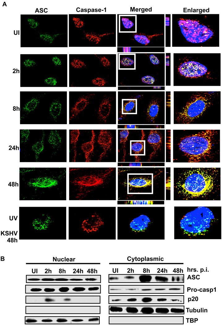
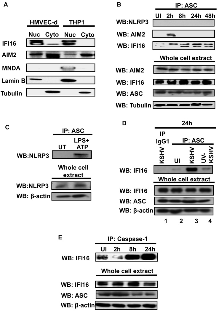
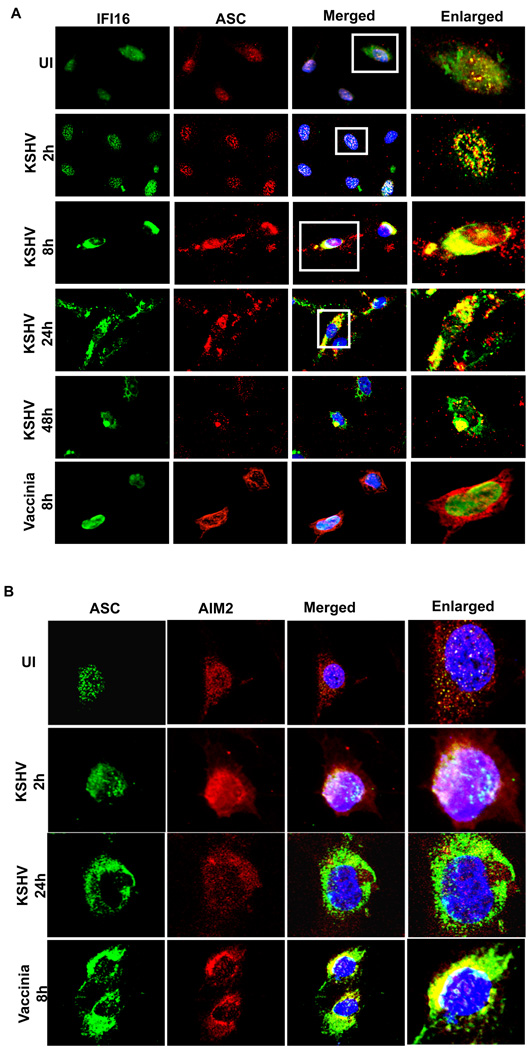
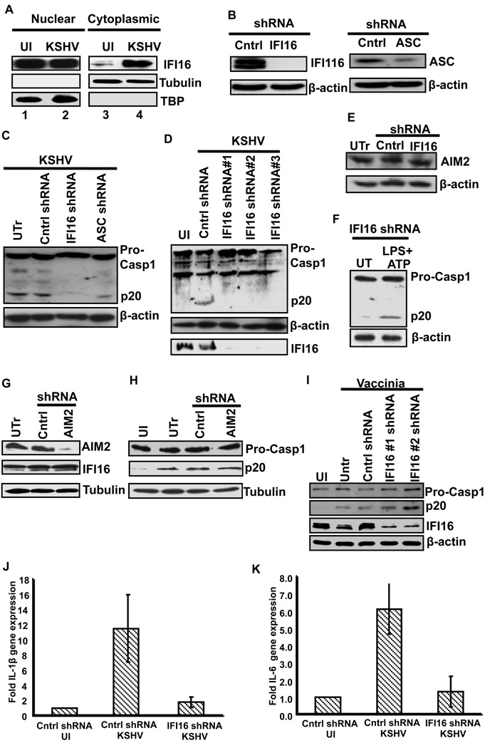
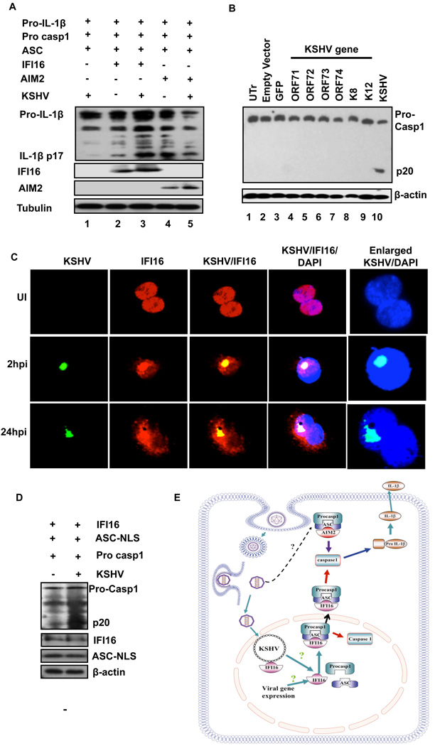
Inflammasomes are cytoplasmic sensors of foreign molecules, including pathogens, and function to induce caspase-1 activation and IL-1β cytokine maturation. Whether such a mechanism exists in the nucleus and is effective against nuclear replicating pathogens is unknown. Nuclear replicating herpesvirus KSHV is associated with Kaposi Sarcoma, an angioproliferative tumor characterized by an inflammatory microenvironment including IL-1β. We demonstrate that during KSHV infection of endothelial cells, interferon gamma-inducible protein 16 (IFI16) interacts with the adaptor molecule ASC and procaspase-1 to form a functional inflammasome. This complex was initially detected in the nucleus and subsequently in the perinuclear area. KSHV gene expression and/or latent KSHV genome is required for inflammasome activation and IFI16 colocalizes with the KSHV genome in the infected cell nucleus. Caspase-1 activation by KSHV was reduced by IFI16 and ASC silencing. Our studies reveal IFI16 as a nuclear pathogen sensor and demonstrate that the inflammasome also functions in the nucleus.


Figures

Figure 1
Figure 1. KSHV infection induces IL-1β maturation and caspase-1 activation
(A) HMVEC-d cells were infected with KSHV (30 DNA copies/cell) and IL-1β gene expression was examined by real-time PCR. Each bar represents the fold increase in gene expression ± SD of three independent experiments. These fold changes were calculated after normalizing with expression of the 18s rRNA gene (B) HMVEC-d cells were infected with KSHV (50 DNA copies/cell) and culture supernatants from infected and uninfected (UI) cells were used to quantitate the secreted IL-1β by ELISA. ** indicate p<0.05. (c and d) KSHV infected (2h) HMVEC-d cells were washed to remove uninternalized virus and cells were either harvested immediately (2h) or incubated for different time points. Proteins were analyzed by immunoblotting for pro- and mature-IL-1β (C), and for pro-caspase-1 and activated caspase-1 (p20) (D). The bands were analyzed and the fold changes in the mature IL-1β and activated caspase-1 levels were calculated considering uninfected as 1 with β-actin as loading control. (E) HMVEC-d cells were uninfected or infected with either live KSHV or UV inactivated KSHV (UV-KSHV) for 2h, washed, harvested immediately (2h) or incubated for 48h. Caspase-1 activation was analyzed as described above.
Figure 2
Figure 2. Inflammasome activation and sub-cellular redistribution of ASC and caspase-1
(A) HMVEC-d cells were uninfected (UI) or infected with either live KSHV or UV-KSHV for 2 h, washed, incubated at 37°C for the indicated time, washed, fixed in 2% paraformaldehyde for 10 min, permeabilized with 0.2% Triton X-100 for 5 min and blocked with signal enhancer. Cells were reacted with anti-ASC and anti-caspase-1 antibodies, washed and incubated with Alexa-488 (green) and Alexa-594 (red) secondary antibodies, respectively. Cell nuclei were visualized by DAPI (blue). The boxed areas were enlarged and shown in the far right panel. (B) HMVEC-d cells were infected with KSHV (30 DNA copies/cell) for 2h, washed and incubated at 37°C for the indicated time. Nuclear and cytoplasmic extracts were examined by immunoblotting with anti-ASC and anti-caspase-1 antibodies. These membranes were stripped and immunoblotted with anti-tubulin and anti-TATA binding protein (TBP) antibodies to check the purity of cytoplasmic and nuclear lysates, respectively and to confirm equal loading.
Figure 3
Figure 3. IFI16 interacts with ASC and caspase-1 in KSHV infected HMVEC-d cells
(A) Expression and sub-cellular distribution of IFI16, AIM2 and MNDA proteins was examined in nuclear and cytoplasmic fractions of uninfected HMVEC-d and THP1 cells by Western blotting. Lamin-B and β-actin show purity and equal loading of nuclear and cytoplasmic fractions, respectively. (B) HMVEC-d cells were infected with KSHV (30 DNA copies/cell) for 2h, washed, and incubated at 37°C for the indicated time. Protein lysates from uninfected (UI) and infected cells were immunoprecipitated with anti-ASC antibodies and Western blotted for NLRP3, AIM2 and IFI16 proteins. Whole cell extracts were blotted with AIM2, IFI16, ASC and tubulin antibodies. (C) HMVEC-d cells were either left untreated (UT) or treated with LPS (20ng/ml for 6h) followed by ATP (5mM for 20min). Protein lysates were immunoprecipitated with anti-ASC antibody and Western blotted for NLRP3. Whole cell extracts were blotted with NLRP3 and b-actin antibodies. (D) HMVEC-d cells were uninfected (UI) or infected with either live KSHV or UV-KSHV for 2h, washed and incubated for 24 h. Protein lysates were immunoprecipitated with anti-ASC antibodies or with irrelevant mouse IgG1 and immunoblotted with anti-IFI16 antibody. (E) Immunoprecipitates prepared by anti-caspase-1 antibodies from uninfected (UI) and KSHV infected HMVEC-d cells were examined for IFI16 by Western blotting. Whole cell extracts were immunoblotted with IFI16, ASC and β-actin antibodies.
Figure 4
Figure 4. Immunofluorescence microscopic analysis of ASC-IFI16 and ASC-AIM2 association in KSHV infected cells
HMVEC-d cells were infected with KSHV (30 DNA copies per cell) or vaccinia virus (5 pfu) for 2h, washed, and incubated at 37°C. At the indicated time, cells were washed, fixed, permeabilized, blocked with signal enhancer, reacted with (A) anti-IFI16 and ASC antibodies and (B) anti-ASC and AIM2 antibodies, washed and incubated with Alexa-488 (green) and Alexa-594 (red) secondary antibodies. The images were merged with DAPI stained nuclei. The boxed areas in Fig. 4a were enlarged without DAPI for clarity (right most panels).
Figure 5
Figure 5. IFI16 undergoes nuclear to cytoplasmic redistribution upon KSHV infection and IFI16 is required for inflammsome activation by KSHV
(A) HMVEC-d cells were infected with KSHV (30 DNA copies/cell) for 2h, washed and infected and uninfected (UI) cells incubated at 37°C for 48h. Nuclear and cytoplasmic extracts were examined by immunobotting with anti-IFI16 antibodies. The purity and equal loading were determined by immunoblotting for TBP and tubulin. (B) HMVEC-d cells were transduced with control shRNA, ASC or IFI16 specific shRNA lentivirus particles and selected with puromycin. Knockdown of IFI16 and ASC protein expression was examined by Western blotting with mouse anti-IFI16 and ASC monoclonal antibodies. (C) Untransduced (UTr), control shRNA (Cntrl) and IFI16 (IFI16 shRNA) and ASC (ASC shRNA) knockdown HMVEC-d cells were infected with KSHV (30 DNA copies/cell) for 2h, washed, incubated at 37°C for 24h, and lysates were analyzed by Western blotting to detect pro-caspase-1 and activated caspase-1 (p20). (D) HMVEC-d cells transduced with control shRNA (Cntrl) or three different IFI16 shRNA lentiviruses were infected with KSHV (30 DNA copies/cell) for 2h, washed, incubated for 24h and lysates were analyzed by Western blotting to detect pro-caspase-1, activated caspase-1 (p20) and IFI16. (E) Protein lysates from untransduced (UTr), control (Cntrl) and IFI16 shRNA transduced HMVEC-d cells were immunoblotted to examine the effect of IFI16 shRNA on AIM2 expression. (F) IFI16 knockdown HMVEC-d were either left untreated (UT) or treated with LPS (20ng/ml for 6h) followed by ATP (5mM for 20min). Protein lysates from untreated and treated cells were immunoblotted with caspase-1 antibody to examine caspase-1 activation. (G) HMVEC-d cells left untransduced (UTr) or transduced with control shRNA (Cntrl) or AIM2 specific shRNA lentivirus particles. The transduced cells were selected with puromycin and then AIM2 and IFI16 protein expression was examined by Western blotting with antibodies against AIM2 and IFI16. (H) Untransduced (UTr), control (Cntrl) and AIM2 knockdown HMVEC-d cells were infected with KSHV (30 DNA copies/cell) for 2h, washed and infected and uninfected (UI) cells further incubated for 24h and lysates were analyzed by Western blotting to detect pro-caspase-1 and activated caspase-1 (p20). (I) Caspase-1 activation was examined in IFI16 knockdown HMVEC-d cells infected with vaccinia virus (5 pfu for 8h) (j and k) Control (cntrl) and IFI16 knockdown cells were infected with KSHV (30 DNA copies/cell) for 2h, washed and infected and uninfected cells incubated for 24h. IL-1β (J) and IL-6 (K) gene expression was examined by real-time RT-PCR. Each bar represents the fold increase in gene expression (compared to uninfected) ± SD of three independent experiments. These fold changes were calculated after normalizing with expression of the18s rRNA gene
Figure 6
Figure 6
(A) Reconstitution of the IFI16 inflammasome. HEK293T cells were cotransfected with 0.5µg of pro-IL-1β, pro-caspase-1, ASC and IFI16 or AIM2 expressing plasmids as indicated. 24h post-transfection, the cells were infected with KSHV (30 DNA copies/cell) for 2h, washed and incubated for 24h. Protein lysates were examined for IL-1β maturation by immunoblotting. Tubulin was used as loading control. (B) Effect of KSHV genes on inflammasome induction. Empty vector, GFP or indicated KSHV genes were expressed in HMVEC-d cells via a lentivirus gene delivery system. Cells were also infected with KSHV for 2 h, washed and kept for 24h. Untransduced (UTr), transduced or KSHV infected cell lysates were examined for caspase-1 activation by immunoblotting with anti-caspase-1 antibodies. Actin was used as loading control. (C) FISH showing IFI16 and KSHV genome interaction. HMVEC-d cells were infected with KSHV (30 DNA copies/cell) for 2h, washed and incubated for the indicated time points. The cells were fixed, permeabilized and immuno-stained with mouse anti-IFI16 antibodies followed by donkey anti-mouse Alexa Fluor 594 secondary antibodies. The cells were then subjected to in situ hybridization with a spectrum green labeled whole KSHV genome probe. (D) HEK293T cells were cotransfected with 0.5mg pro-caspase-1, IFI16 and ASC-NLS expressing plasmids as indicated. 24h post-transfection, the cells were infected with KSHV (30 DNA copies/ cell) for 2h, washed, incubated for 24h and lysates were examined for caspase-1 activation by immunoblotting. (E) Schematic depicting a model for IFI16 mediated inflammasome activation in response to KSHV infection of endothelial cells. In uninfected endothelial cells, IFI16, ASC and caspase-1 are predominantly distributed in the nucleus. During de novo infection leading to the establishment of latency, IFI16, ASC and procaspase-1 form the multi-protein inflammasome complex in the nucleus leading to activation of caspase-1. The IFI16 containing inflammasome complex also migrates from the nucleus to the cytoplasm and forms peri-nuclear aggregates. Early during de novo infection, KSHV also induces the interaction between ASC and AIM2 possibly suggesting that the AIM2 inflammasome might be involved in sensing during the virus internalization process.

Comment in

Similar articles

Cited by

References

    1. Aglipay JA, Lee SW, Okada S, Fujiuchi N, Ohtsuka T, Kwak JC, Wang Y, Johnstone RW, Deng C, Qin J, et al. A member of the Pyrin family, IFI16, is a novel BRCA1-associated protein involved in the p53-mediated apoptosis pathway. Oncogene. 2003;22:8931–8938. - PubMed
    1. Bryan NB, Dorfleutner A, Kramer SJ, Yun C, Rojanasakul Y, Stehlik C. Differential splicing of the apoptosis-associated speck like protein containing a caspase recruitment domain (ASC) regulates inflammasomes. J Inflamm (Lond) 2010;7:23. - PMC - PubMed
    1. Bryan NB, Dorfleutner A, Rojanasakul Y, Stehlik C. Activation of inflammasomes requires intracellular redistribution of the apoptotic speck-like protein containing a caspase recruitment domain. J Immunol. 2009;182:3173–3182. - PMC - PubMed
    1. Burckstummer T, Baumann C, Bluml S, Dixit E, Durnberger G, Jahn H, Planyavsky M, Bilban M, Colinge J, Bennett KL, et al. An orthogonal proteomic-genomic screen identifies AIM2 as a cytoplasmic DNA sensor for the inflammasome. Nat Immunol. 2009;10:266–272. - PubMed
    1. Case CL, Shin S, Roy CR. Asc and Ipaf Inflammasomes direct distinct pathways for caspase-1 activation in response to Legionella pneumophila. Infect Immun. 2009;77:1981–1991. - PMC - PubMed

Publication types