. 2011 May 15;71(10):3688-700.
doi: 10.1158/0008-5472.CAN-10-3445.

ΔNp63 versatilely regulates a Broad NF-κB gene program and promotes squamous epithelial proliferation, migration, and inflammation

Affiliations

ΔNp63 versatilely regulates a Broad NF-κB gene program and promotes squamous epithelial proliferation, migration, and inflammation

Xinping Yang et al. Cancer Res. .

Erratum in

  • Cancer Res. 2011 Dec 1;71(23):7323

Abstract

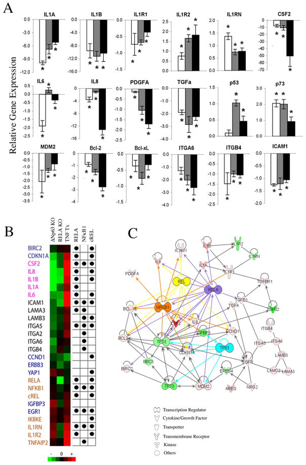
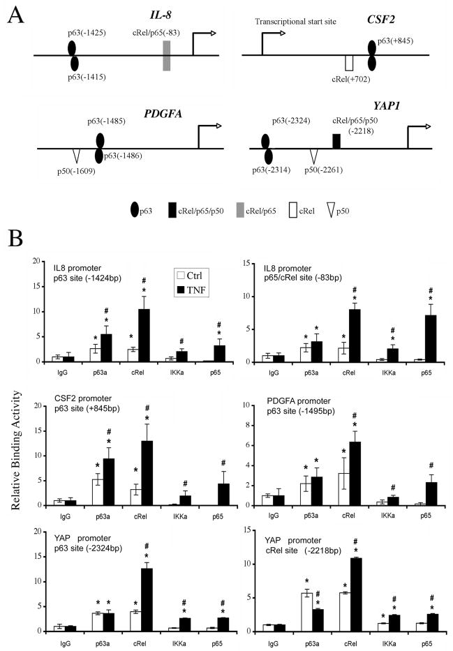
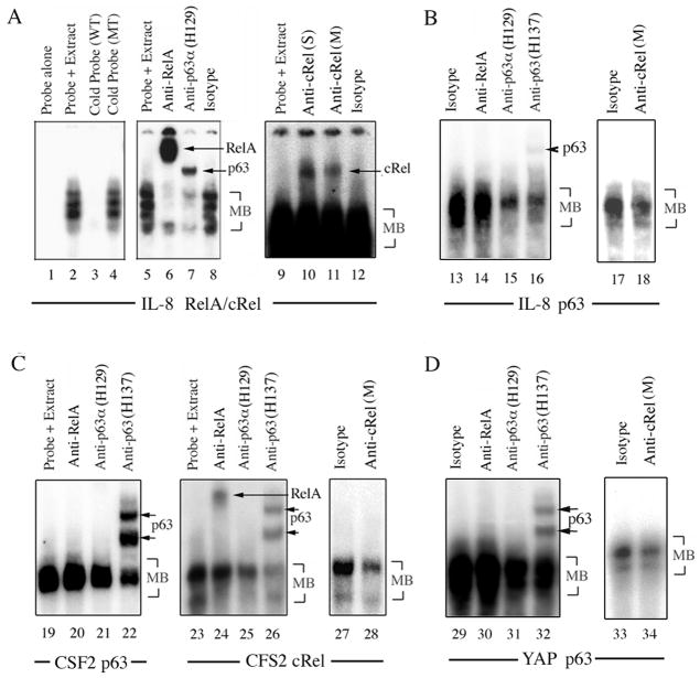
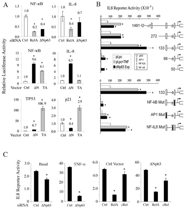
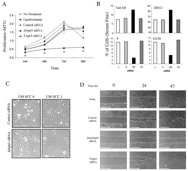
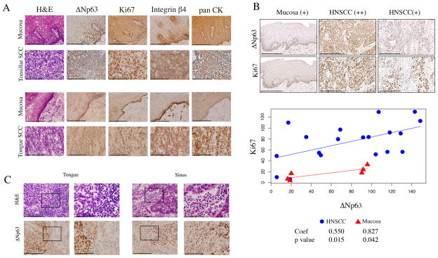
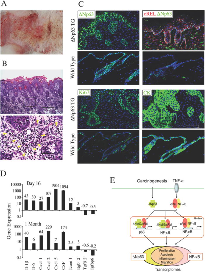
Head and neck squamous cell carcinoma (HNSCC) and many other epithelial malignancies exhibit increased proliferation, invasion, and inflammation, concomitant with aberrant nuclear activation of TP53 and NF-κB family members ΔNp63, cRel, and RelA. However, the mechanisms of cross-talk by which these transcription factors coordinate gene expression and the malignant phenotype remain elusive. In this study, we showed that ΔNp63 regulates a cohort of genes involved in cell growth, survival, adhesion, and inflammation, which substantially overlaps with the NF-κB transcriptome. ΔNp63 with cRel and/or RelA are recruited to form novel binding complexes on p63 or NF-κB/Rel sites of multitarget gene promoters. Overexpressed ΔNp63- or TNF-α-induced NF-κB and inflammatory cytokine interleukin-8 (IL-8) reporter activation depended on RelA/cRel regulatory binding sites. Depletion of RelA or ΔNp63 by small interfering RNA (siRNA) significantly inhibited NF-κB-specific, or TNF-α-induced IL-8 reporter activation. ΔNp63 siRNA significantly inhibited proliferation, survival, and migration by HNSCC cells in vitro. Consistent with these data, an increase in nuclear ΔNp63, accompanied by increased proliferation (Ki-67) and adhesion (β4 integrin) markers, and induced inflammatory cell infiltration was observed throughout HNSCC specimens, when compared with the basilar pattern of protein expression and minimal inflammation seen in nonmalignant mucosa. Furthermore, overexpression of ΔNp63α in squamous epithelial cells in transgenic mice leads to increased suprabasilar cRel, Ki-67, and cytokine expression, together with epidermal hyperplasia and diffuse inflammation, similar to HNSCC. Our study reveals ΔNp63 as a master transcription factor that, in coordination with NF-κB/Rels, orchestrates a broad gene program promoting epidermal hyperplasia, inflammation, and the malignant phenotype of HNSCC.


Conflict of interest statement

Disclosure of Conflicts of Interest: No conflicts of interest were identified.

Figures

Figure 1
Figure 1. Knockdown of ΔNp63 regulates a broad gene expression program that overlaps with the NF-κB transcriptome and network
A, Relative fold-change in mRNA expression after ΔNp63 siRNA transfection of UM-SCC 1 for 24, 48 or 72 hrs (white, grey, black bars); *p<0.05 vs. control siRNA. B, Heat map for up- (red) or down-regulated (green) mRNA expression after ΔNp63, RELA siRNA or TNF-α treatment. Dots indicate predicted RELA, NF-κB1 and c-REL binding sites in the proximal promoter regions (−500bp ~ +100bp of TSS). Gene categories are: Blue, growth and apoptosis; pink, cytokines; black, adhesion; brown, NF-κB. C, Ingenuity Pathway Analysis (IPA) illustrates important functional networks between ΔNp63-modulated genes and RELA, c-REL or NFκB1.
Figure 2
Figure 2. Distinct ΔNp63, cREL and RELA binding activities on p63 or NF-κB responsive elements
A, p63 and NF-κB/REL binding sites (with base pairs from transcription start site) on gene promoters were predicted by Genomatix by including user-defined p63 PWM as described in Supplemental Methods. B, ChIP assay performed using anti-p63, c-REL, IKKα, RELA and isotype antibodies without (Ctrl) or with TNF-α treatment, followed by real-time PCR. Mean Relative binding activity +/−SD for triplicates; p<0.05 compared with isotype (*), or between Ctrl and TNF-α treatment (#).
Figure 3
Figure 3. Coordinate binding of p63, c-REL and/or RELA protein complexes to p63 and NF-κB regulatory elements
EMSA using nuclear extract from UM-SCC1 treated with TNF-α, incubated with 32P-labeled oligonucleotide probes for A, known IL-8 promoter RELA/cREL (−83bp, lane 1–12), and B, predicted p63 (−1425bp, lane 13–18) sites; C, CSF2 promoter p63 (+845bp, lane 19–22) or c-REL (+702bp, lane 23–28) sites; and D, a YAP promoter p63 site (−2324bp, lane 29–34). Experimental conditions include labeled probe alone; labeled probes plus nuclear extracts; labeled probe and nuclear extract plus 50-fold excess unlabeled wild-type (WT) or mutant (MT) competitor DNA; anti-RELA, anti-p63 (H129 or H137), anti-c-REL (S: Santa Cruz; M: Millipore), or isotype control antibodies. MB: main band.
Figure 4
Figure 4. ΔNp63 and TNF-α modulate NF-κB, IL-8, TP53 and p21 reporter activities
A, Relative NF-κB-specific, NF-κB-dependent IL-8 (−133bp), TP53-specific and TP53-dependent p21Cip1 luciferse reporter activities 48 hours after transfection of UM-SCC1 with Ctrl, RELA or Δ Np63 siRNAs, or overexpression of Ctrl, ΔNp63α or TAp63α vectors, as indicated. Normalized with β-gal. *p<0.05 vs. Ctrl. B, Relative IL-8 luciferase reporter activity for serially deleted or binding site-specific point mutant promoter constructs, which include p63, NF-κB, AP-1, and NF-IL6 binding sites. UM-SCC1 cells were transfected with indicated reporter constructs plus Ctrl (pLpc), TNF-α (20ng/ml), or ΔNp63α expression vectors. Mean+SD of reporter activities of 6 replicates, adjusted for cell density by WST1 48 hrs after transfection. *p<0.05 vs. baseline. C, IL-8 promoter (−133bp) reporter activity; Left panels, Basal (without) or with TNF-α for 24 hours preceded by Ctrl or ΔNp63 siRNA for 46 hours. Right panels, co-transfected with Ctrl or ΔNp63 expression vector, and Ctrl, RELA or c-RELs siRNAs for 70 hours. Mean+SD for four replicates, after normalization for cell density by WST1. *p<0.05 vs. ctrl.
Figure 5
Figure 5. ΔNp63 regulates HNSCC proliferation, cell cycle, differentiation and migration
UM-SCC1 cells were transfected with lipofectamine (L), control (C), ΔNp63 or TAp63 siRNA as indicated. A, Cell proliferation measured by MTT assay. *p<0.05 vs control. B, DNA cell cycle distribution (G0/G1-S-G2/M) at 48 hrs and DNA fragmentation (sub G0) measured at 72hrs by flow cytometry. C, Photomicrographs of cell morphology after 96 hours (100X, scale bar: 100μm). D, Wound assay, UM-SCC1 monolayers were scratched 24 hrs after indicated transfections, and wound closure was photographed and measured at indicated time points (40X, scale bar: 1000μm).
Figure 6
Figure 6. Diffuse ΔNp63, Ki67, and integrin β4 staining with inflammation in HNSCC compared to basilar staining pattern and lack of inflammation in mucosa
A, frozen sections of tonsillar and tongue SCC with matched mucosa were stained with H&E, and ΔNp63, Ki67, Integrin β4, and pan CK antibodies by immunoperoxidase (400X, scale bar: 200μM). B, tissue array of HNSCC was immunostained and quantified using histoscore system (200X, scale bar: 200μM). Linear regression and Pearson correlation for ΔNp63 and Ki67. Coefficient of the slope and statistical significance are shown. C, ΔNp63 staining in a tongue and sinus HNSCC, with adjacent infiltrating inflammatory cells. Left panels: 400X, scale bar: 200μM. The black rectangle frames highlight the areas shown at 1000X in right panels, scale bar: 80μM.
Figure 7
Figure 7. Skin from ΔNp63α transgenic mice demonstrates hyperplasia, cellular inflammation, and increased proinflammatory cytokine expression
ΔNp63α was induced in skin of TG animals for two months after birth. A, gross morphology, erythematous lesions. B, Histology, H&E stain. Upper panel, epidermal hyperplasia and cellular inflammation in dorsal skin sections (100X, scale bar: 300μm); Lower panel, infiltrating inflammatory cells in the dermis highlighted by yellow arrowheads (600X, scale bar 50μm). C, dorsal skin sections from ΔNp63α TG and wild-type mice were stained with anti-ΔNp63, cRel, Ki67, and K14 (CK) antibodies and DAPI counterstain (scale bar: 75μm). D, gene expression in dorsal skin 16 days (upper), or 1 month (bottom) following ΔNp63α induction in neonatal TG mice, vs. wild-type littermates. E, Model of ΔNp63 and NF-κB/REL interactions mediating regulation of a broad gene program. Overexpressed ΔNp63 following carcinogenesis, and c-REL or RELA activated by TNF-α from inflammation in the tumor microenvironment, interact via p63 or NF-κB sequences of target gene promoters. p63/REL protein complexes regulate a broad gene program that overlaps with NF-κB transcriptome and promotes the malignant phenotype.

Similar articles

Cited by

References

    1. Karin M. Nuclear factor-kappaB in cancer development and progression. Nature. 2006;441:431–6. - PubMed
    1. Stiewe T. The p53 family in differentiation and tumorigenesis. Nat Rev Cancer. 2007;7:165–8. - PubMed
    1. Dey A, Tergaonkar V, Lane DP. Double-edged swords as cancer therapeutics: simultaneously targeting p53 and NF-kappaB pathways. Nat Rev Drug Discov. 2008;7:1031–40. - PubMed
    1. Van Waes C. Nuclear factor-kappaB in development, prevention, and therapy of cancer. Clin Cancer Res. 2007;13:1076–82. - PubMed
    1. Tergaonkar V, Pando M, Vafa O, Wahl G, Verma I. p53 stabilization is decreased upon NFkappaB activation: a role for NFkappaB in acquisition of resistance to chemotherapy. Cancer Cell. 2002;1:493–503. - PubMed

Publication types

MeSH terms