. 2011 Jul 8;286(27):24374-84.
doi: 10.1074/jbc.M111.230797. Epub 2011 May 16.

The interaction between two extracellular linker regions controls sustained opening of acid-sensing ion channel 1

Affiliations

The interaction between two extracellular linker regions controls sustained opening of acid-sensing ion channel 1

Andreas Springauf et al. J Biol Chem. .

Abstract

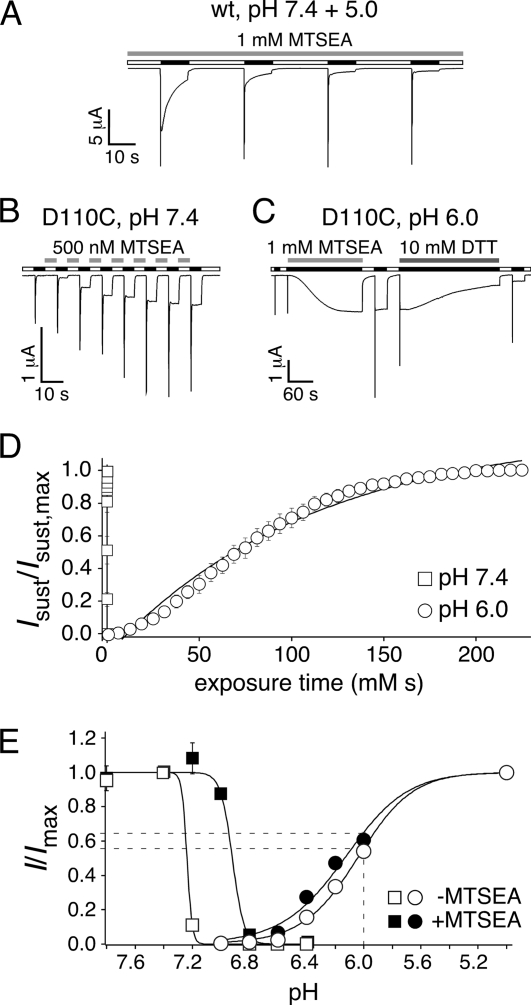
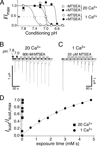
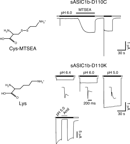
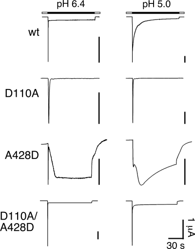
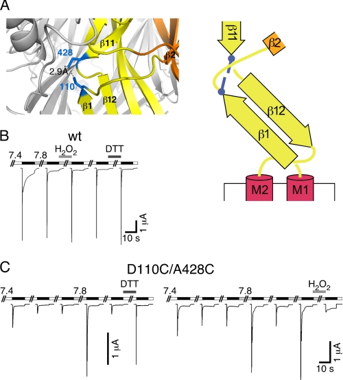
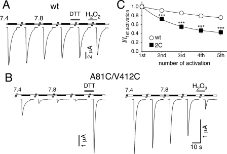
Activation of acid-sensing ion channels (ASICs) contributes to neuronal death during stroke, to axonal degeneration during neuroinflammation, and to pain during inflammation. Although understanding ASIC gating may help to modulate ASIC activity during these pathologic situations, at present it is poorly understood. The ligand, H(+), probably binds to several sites, among them amino acids within the large extracellular domain. The extracellular domain is linked to the two transmembrane domains by the wrist region that is connected to two anti-parallel β-strands, β1 and β12. Thus, the wrist region together with those β-strands may have a crucial role in transmitting ligand binding to pore opening and closing. Here we show that amino acids in the β1-β2 linker determine constitutive opening of ASIC1b from shark. The most crucial residue within the β1-β2 linker (Asp(110)), when mutated from aspartate to cysteine, can be altered by cysteine-modifying reagents much more readily when channels are closed than when they are desensitized. Finally, engineering of a cysteine at position 110 and at an adjacent position in the β11-β12 linker leads to spontaneous formation of a disulfide bond that traps the channel in the desensitized conformation. Collectively, our results suggest that the β1-β2 and β11-β12 linkers are dynamic during gating and tightly appose to each other during desensitization gating. Hindrance of this tight apposition leads to reopening of the channel. It follows that the β1-β2 and β11-β12 linkers modulate gating movements of ASIC1 and may thus be drug targets to modulate ASIC activity.


Figures

FIGURE 1.
FIGURE 1.
Representative current traces for rASIC1a (top panels) and sASIC1b (bottom panels) illustrating the different desensitization kinetics at mild and strong acidification (pH 6.4 and 5.0). The time constant of desensitization of transient rASIC1a currents was τ = 2.1 ± 0.2 s at pH 6.4 (n = 13) and τ = 1.8 ± 0.2 s at pH 5.0 (n = 13; p = 0.1). Desensitization was complete, and no sustained current remained. Desensitization of the transient sASIC1b currents was much faster than for rASIC1a (τ < 50 ms, n = 13; p < 0.001) but incomplete. Desensitization of the second current component at pH 5.0 was best described by two time constants (τ1 = 7 ± 0.4 s and τ2 = 2.1 ± 0.4 s; n = 6). Note that current amplitudes at pH 5.0 were larger than at pH 6.4.
FIGURE 2.
FIGURE 2.
A small region shortly after TMD1 determines sustained opening of sASIC1b. Left panel, schematic drawings of rASIC1a and sASIC1b and chimeras. Middle panel, representative traces of currents at pH 6.4 and 5.0. The scale bars correspond to 2 μA, except when otherwise indicated. Right panel, time constants of desensitization of the transient current at pH 6.4 (open circles) and 5 (filled circles). n > 6. ***, p < 0.001 (compared with sASIC1b).
FIGURE 3.
FIGURE 3.
Residues 109–115 of sASIC1b localize to the β1-β2 linker. A, ribbon representation of the desensitized cASIC1 structure. The different domains of the ECD are shown in different colors (TMDs in red, palm in yellow, thumb in green, knuckle in turquoise, finger in purple, and β-ball in orange). B, β-strands 1 and 12 from one subunit and the linkers that connect them to β-strands 2 and 11, respectively, are shown. C, schematic representation of B. D, amino acid sequences of the β1-β2 and the β11-β12 linkers of sASIC1b, rASIC1a, and cASIC1, respectively. The carboxyl-carboxylate pair between β-strands 1 and 12 is illustrated by a red line.
FIGURE 4.
FIGURE 4.
Aspartate 110 is most crucial for sustained opening of sASIC1b. A and B, left panels, representative current traces at pH 6.4 and 5.0 for sASIC1b (A), rASIC1a (B), and amino acid substitutions in the β1-β2 linker. The scale bars correspond to 2 μA, except when otherwise indicated. The insets show the current decline after washout of acidic pH on a 4-fold expanded scale. Right panels, time constants of desensitization of the transient current at pH 6.4 (open circles) and 5 (filled circles). n > 8. *, p < 0.05; ***, p < 0.001 (compared with wild type). C, left panel, current-voltage relationship for the transient and the sustained current of rASIC1a-MDS at pH 5.0. Channels had been repeatedly activated at different holding potentials and currents normalized to the current at −70 mV. The absolute values of the current amplitudes at −70 mV were 7.2 ± 1.7 μA (transient current; n = 11), and 0.77 ± 0.13 μA (sustained current; n = 10), respectively. The current-voltage relationship for voltages between −30 and +50 mV for rASIC1a-wt is shown for comparison as dotted squares; currents were normalized to the current at −30 mV (n = 3). Right panel, representative current trace illustrating constitutive activity of rASIC1a-MDS. Amiloride (1 mm) and substitution of Na+ by the large cation NMDG+ reduced the background current, revealing some constitutive activity of rASIC1a-MDS at pH 7.4.
FIGURE 5.
FIGURE 5.
MTS modification of sASIC1b-D110C leads to sustained opening. A, 1 mm MTSEA, when applied at pH 7.4 and 5.0, did not change the current of wt-sASIC1b. B, 500 nm MTSEA, applied for 5 s in the interval between activation (at pH 7.4), induced robust sustained currents for sASIC1b-D110C. Activation was with pH 6 for 5 s. C, a higher concentration of MTSEA (1 mm) than in B, applied at pH 6 when channels were desensitized, also induced robust sustained currents. Note also the different time scales in B and C. The sustained currents could be reversed by application of 10 mm DTT. D, increase in sustained currents of sASIC1b-D110C as a function of exposure time (time exposed × concentration MTSEA) when MTSEA was applied at pH 7.4 (open squares) or at pH 6 (open circles), in experiments like those shown in B and C. The symbols represent the mean amplitudes of the sustained currents, normalized to the maximal sustained current, and quantified every 5 s (n = 8); the lines represent exponential fits. E, activation curves (circles) and SSD curves (squares) for peak currents of sASIC1b-D110C before (open symbols) and after MTSEA modification (100 μm for 60 s at pH 7.4; filled symbols). For activation curves, the channels were activated with different acidic solutions, as indicated, from a conditioning pH 7.4 (n = 8, without MTSEA; n = 9, with MTSEA). For SSD curves, channels were activated with pH 5 with different conditioning pH, as indicated (n = 8, without MTSEA; n = 6, with MTSEA). Peak current amplitudes were normalized to the peak current amplitude at pH 5.0 (activation curves) or with conditioning pH 7.4 (SSD curves), respectively. The dotted lines illustrate the expected increase in peak current at pH 6 by MTS modification, which is too small to explain the increase in peak current amplitude observed in experiments like in B and C.
FIGURE 6.
FIGURE 6.
MTS modification of sASIC1b-D110C is state-dependent. A, SSD curves for peak currents of sASIC1b-D110C in 1 mm Ca2+ (circles) and 20 mm Ca2+ (squares) before (open symbols) and after MTSEA modification (100 μm for 60 s at pH 7.4; filled symbols). The channels were activated with pH 5 with different conditioning pH levels, as indicated (n = 8). Peak current amplitudes were normalized to the peak current amplitude with conditioning pH 7.8 (1 mm Ca2+) or pH 7.4 (20 mm Ca2+), respectively. The dotted line illustrates the partial desensitization at pH 7 and in 20 mm Ca2+ (before MTS modification). B, 600 nm MTSEA was applied in 20 mm Ca2+ for 10 s in the interval between activation (pH 6, 5 s) of the channel. C, a higher concentration of MTSEA (20 μm) than in B was applied in 1 mm Ca2+ for 30 s in the interval between activation (pH 6, 5 s) of the channel. Note also the different time scales in B and C. D, increase in sustained currents as a function of exposure time (time exposed × concentration MTSEA) when MTSEA was applied in 20 mm Ca2+ (closed squares) or in 1 mm Ca2+ (closed circles) in experiments like those shown in B and C. For both Ca2+ concentrations, conditioning pH was 7.0. The symbols represent the mean amplitudes of the sustained currents (sust), normalized to the maximal sustained (sust,max) current and quantified every 30 s with 1 mm Ca2+ and every 10 s with 20 mm Ca2+ (n = 4); the lines represent exponential fits.
FIGURE 7.
FIGURE 7.
A Lys at position 110 mimics MTSEA modification. Left panels, chemical structures of an MTSEA-modified cysteine and a lysine. Right panels, representative current traces for MTSEA modification of sASIC1b-D110C (top panel) and for sASIC1b-D110K (bottom panel). The currents for sASIC1b-D110K are shown at three different pH values. The current rise time is also shown on an expanded time scale. The current trace at the bottom illustrates amiloride sensitivity (1 mm) of the sustained sASIC1b-D110K current.
FIGURE 8.
FIGURE 8.
Representative current traces at pH 6.4 and 5.0 for sASIC1b-wt, -D110A, -A428D, and -D110A/A428D, respectively. The D110A substitution abolished sustained currents, whereas the A428D substitution strongly increased sustained currents. Combined substitutions (D110A/A428D) conferred sustained currents that resembled those of wild type.
FIGURE 9.
FIGURE 9.
Cross-linking of residues 110 and 428 traps sASIC1b in the desensitized state. A, left panel, detail from the cASIC1 crystal structure, in which two Cys residues had been modeled: one at position 82 and one at position 413 (corresponding to positions 110 and 428 of sASIC1b). Right panel, schematic representation with the two cysteines as blue bars. B, reducing and oxidizing conditions had no effect on sASIC1b-wt currents. C, left panel, sASIC1b-D110C/A428C current amplitude gradually decreased with repeated stimulation by ligand (pH 5.0). Switching the pH to 7.8 or reducing conditions strongly increased current amplitude. Right panel, oxidizing conditions strongly reduced current amplitude of sASIC1b-D110C/A428C.
FIGURE 10.
FIGURE 10.
Cross-linking of residues 81 and 412 traps rASIC1a in the desensitized state. A, reducing and oxidizing conditions had no effect on rASIC1a-wt currents. B, left panel, rASIC1a-A81C/V412C current amplitude gradually decreases with repeated stimulation by ligand (pH 6.4). Switching the pH to 7.8 has no effect on current amplitude, whereas reducing conditions dramatically increased current amplitude. Right panel, oxidizing conditions reduced current amplitude of rASIC1a-A81C/V412C. C, quantification of tachyphylaxis for rASIC1a-wt and -A81C/V412C (2C). The channels were repeatedly activated, and current amplitudes were normalized to the first amplitude. Repeated activation reduced rASIC1a-A81C/V412C currents significantly more strongly than wt currents. Absolute values of the initial amplitudes were 14.3 ± 2.5 μA (wt; n = 6) and 1.0 ± 0.2 μA (A81C/V412C; n = 6), respectively. The lines represent fits to a mono-exponential function. ***, p < 0.001.

References

    1. Wemmie J. A., Price M. P., Welsh M. J. (2006) Trends Neurosci. 29, 578–586 - PubMed
    1. Baron A., Waldmann R., Lazdunski M. (2002) J. Physiol. 539, 485–494 - PMC - PubMed
    1. Askwith C. C., Wemmie J. A., Price M. P., Rokhlina T., Welsh M. J. (2004) J. Biol. Chem. 279, 18296–18305 - PubMed
    1. Xiong Z. G., Zhu X. M., Chu X. P., Minami M., Hey J., Wei W. L., MacDonald J. F., Wemmie J. A., Price M. P., Welsh M. J., Simon R. P. (2004) Cell 118, 687–698 - PubMed
    1. Wemmie J. A., Chen J., Askwith C. C., Hruska-Hageman A. M., Price M. P., Nolan B. C., Yoder P. G., Lamani E., Hoshi T., Freeman J. H., Jr., Welsh M. J. (2002) Neuron 34, 463–477 - PubMed

Publication types