

Decoding the membrane activity of the cyclotide kalata B1: the importance of phosphatidylethanolamine phospholipids and lipid organization on hemolytic and anti-HIV activities
- PMID: 21576247
- PMCID: PMC3129204
- DOI: 10.1074/jbc.M111.253393
Decoding the membrane activity of the cyclotide kalata B1: the importance of phosphatidylethanolamine phospholipids and lipid organization on hemolytic and anti-HIV activities
Abstract
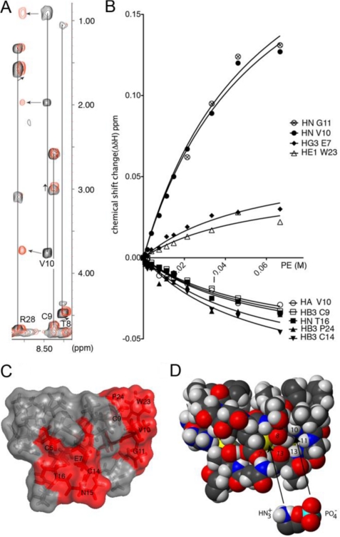
Cyclotides, a large family of cyclic peptides from plants, have a broad range of biological activities, including insecticidal, cytotoxic, and anti-HIV activities. In all of these activities, cell membranes seem likely to be the primary target for cyclotides. However, the mechanistic role of lipid membranes in the activity of cyclotides remains unclear. To determine the role of lipid organization in the activity of the prototypic cyclotide, kalata B1 (kB1), and synthetic analogs, their bioactivities and affinities for model membranes were evaluated. We found that the bioactivity of kB1 is dependent on the lipid composition of target cell membranes. In particular, the activity of kB1 requires specific interactions with phospholipids containing phosphatidylethanolamine (PE) headgroups but is further modulated by nonspecific peptide-lipid hydrophobic interactions, which are favored in raft-like membranes. Negatively charged phospholipids do not favor high kB1 affinity. This lipid selectivity explains trends in antimicrobial and hemolytic activities of kB1; it does not target bacterial cell walls, which are negatively charged and lacking PE-phospholipids but can insert in the membranes of red blood cells, which have a low PE content and raft domains in their outer layer. We further show that the anti-HIV activity of kB1 is the result of its ability to target and disrupt the membranes of HIV particles, which are raft-like membranes very rich in PE-phospholipids.
Figures


Similar articles
-
Molecular dynamics simulations support a preference of cyclotide kalata B1 for phosphatidylethanolamine phospholipids.Biochim Biophys Acta Biomembr. 2024 Mar;1866(3):184268. doi: 10.1016/j.bbamem.2023.184268. Epub 2024 Jan 6. Biochim Biophys Acta Biomembr. 2024. PMID: 38191035 Review.
-
Phosphatidylethanolamine binding is a conserved feature of cyclotide-membrane interactions.J Biol Chem. 2012 Sep 28;287(40):33629-43. doi: 10.1074/jbc.M112.372011. Epub 2012 Aug 1. J Biol Chem. 2012. PMID: 22854971 Free PMC article.
-
Kalata B1 and Kalata B2 Have a Surfactant-Like Activity in Phosphatidylethanolomine-Containing Lipid Membranes.Langmuir. 2017 Jul 5;33(26):6630-6637. doi: 10.1021/acs.langmuir.7b01642. Epub 2017 Jun 21. Langmuir. 2017. PMID: 28605904
-
The self-association of the cyclotide kalata B2 in solution is guided by hydrophobic interactions.Biopolymers. 2013 Sep;100(5):453-60. doi: 10.1002/bip.22269. Biopolymers. 2013. PMID: 23893463
-
Importance of the cell membrane on the mechanism of action of cyclotides.ACS Chem Biol. 2012 Apr 20;7(4):626-36. doi: 10.1021/cb200395f. Epub 2012 Feb 3. ACS Chem Biol. 2012. PMID: 22260456 Review.
Cited by
-
Free ISG15 Inhibits the Replication of Peste des Petits Ruminants Virus by Breaking the Interaction of Nucleoprotein and Phosphoprotein.Microbiol Spectr. 2022 Oct 26;10(5):e0103122. doi: 10.1128/spectrum.01031-22. Epub 2022 Aug 29. Microbiol Spectr. 2022. PMID: 36036587 Free PMC article.
-
Lipid-Centric Approaches in Combating Infectious Diseases: Antibacterials, Antifungals and Antivirals with Lipid-Associated Mechanisms of Action.Antibiotics (Basel). 2023 Dec 11;12(12):1716. doi: 10.3390/antibiotics12121716. Antibiotics (Basel). 2023. PMID: 38136750 Free PMC article. Review.
-
The Activity of Chelidonium majus L. Latex and Its Components on HPV Reveal Insights into the Antiviral Molecular Mechanism.Int J Mol Sci. 2022 Aug 17;23(16):9241. doi: 10.3390/ijms23169241. Int J Mol Sci. 2022. PMID: 36012505 Free PMC article.
-
A Synthetic mirror image of kalata B1 reveals that cyclotide activity is independent of a protein receptor.Chembiochem. 2011 Nov 4;12(16):2456-62. doi: 10.1002/cbic.201100450. Epub 2011 Sep 16. Chembiochem. 2011. PMID: 21928440 Free PMC article.
-
Do plant cyclotides have potential as immunosuppressant peptides?J Nat Prod. 2012 Feb 24;75(2):167-74. doi: 10.1021/np200722w. Epub 2012 Jan 24. J Nat Prod. 2012. PMID: 22272797 Free PMC article.
References
-
- Craik D. J., Daly N. L., Bond T., Waine C. (1999) J. Mol. Biol. 294, 1327–1336 - PubMed
-
- Colgrave M. L., Craik D. J. (2004) Biochemistry 43, 5965–5975 - PubMed
-
- Gran L., Sandberg F., Sletten K. (2000) J. Ethnopharmacol. 70, 197–203 - PubMed
-
- Gustafson K. R., Sowder R. C. I., Henderson L. E., Parsons I. C., Kashman Y., Cardellina J. H. I., McMahon J. B., Buckheit R. W. J., Pannell L. K., Boyd M. R. (1994) J. Am. Chem. Soc. 116, 9337–9338
Publication types
MeSH terms
Substances
LinkOut - more resources
Full Text Sources
Other Literature Sources
Medical
