Review
. 2011 Aug 1;3(8):a004796.
doi: 10.1101/cshperspect.a004796.

Phosphoinositides in cell architecture

Affiliations
Review

Phosphoinositides in cell architecture

Annette Shewan et al. Cold Spring Harb Perspect Biol. .

Abstract

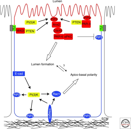
Inositol phospholipids have been implicated in almost all aspects of cellular physiology including spatiotemporal regulation of cellular signaling, acquisition of cellular polarity, specification of membrane identity, cytoskeletal dynamics, and regulation of cellular adhesion, motility, and cytokinesis. In this review, we examine the critical role phosphoinositides play in these processes to execute the establishment and maintenance of cellular architecture. Epithelial tissues perform essential barrier and transport functions in almost all major organs. Key to their development and function is the establishment of epithelial cell polarity. We place a special emphasis on highlighting recent studies demonstrating phosphoinositide regulation of epithelial cell polarity and how individual cells use phosphoinositides to further organize into epithelial tissues.


Figures

Figure 1.
Figure 1.
Phosphoinositide subcellular distribution, metabolism, and protein effectors. (A) Subcellular distribution of PI species. PIs concentrate in cytosolic membranes, serving as discrete determinants of membrane identity. The predominant localization of particular PI species in subcellular compartments is depicted. There is some overlap of PI signature between membrane compartments, and heterogeneity of PI distribution on membrane compartments also occurs. PtdIns(4,5)P2 and PtdIns(3,4,5)P3 are enriched at the plasma membrane, possibly in raft-like domains. PtdIns(3,4)P2 dominates in early endocytic membranes and at the plasma membrane. PtdIns(3)P is concentrated on early endosomal (EE) membranes, and the multivesicular body (MVB) compartment. PtdIns(4)P is enriched at the Golgi complex and in Golgi-derived carriers. PtdIns(3,5)P2 concentrates on late compartments of the endocytic pathway, the MVB, and lysosome. PtdIns(5)P is localized in the nucleus, and generation of nuclear PtdIns(4,5)P2 is key to regulating some aspects of gene expression (reviewed in Barlow et al. 2009). Parallels can be drawn between the generation of front–rear axis in migrating cells and apical–basal polarity in polarized cell types (for recent review, see Nelson 2009). Not all cellular compartments are illustrated and arrows are not intended to represent the entire cohort of known endocytic trafficking routes. Illustration based in part on data from Kutateladze (2010), with additional elements added. (B) Representation of the metabolic interconversions that generate the seven phosphoinositide species from PtdIns. Kinases and phosphatases involved in generating the PIs involved in apical and basolateral membrane identity are indicated. (C) PI binding modules present in cytosolic effectors and their reported PI binding preferences. The family of PI “code-breaking” modules includes PH, ANTH (AP180 amino-terminal homology), C2 (conserved region 2 of protein kinase C), ENTH (epsin amino-terminal homology), FERM (band 4.1, Ezrin, Radixin, Moesin), FYVE (Fab1, YOTB, Vac1, and EEA1), GOLPH3 (Golgi phosphoprotein 3), PROPPINS (B-propellors that bind PIs), PTB (phosphotyrosine binding), PX (Phox homology), and Tubby modules. Examples of protein interactions with different PIs are provided. (Panel based in part on data from DiPaolo and Di Camilli 2006; McCrea and De Camilli 2009; and Kutateladze 2010.)
Figure 2.
Figure 2.
PIs specify membrane identity in epithelial cells. PI(3)-kinase generated PIP3 (PtdIns(3,4,5)P3) at the basolateral surface contributes to apico-basal polarity specification. This is coordinated with PTEN and PI(5)-kinase enrichment of PIP2 (PtdIns(4,5)P2) at the apical surface. A PTEN/PIP2/Anx2/Cdc42 pathway links the production of PIP2 to actin reorganization during apical membrane biogenesis and lumen formation.
Figure 3.
Figure 3.
PI control of actin assembly and organization supports junctional integrity and tissue cohesion. (A) Coordination of RhoGTPase directed Arp2/3-dependent actin assembly supports AJ stability. PI control of RhoGTPase localization and activity directs differential Arp2/3-mediated actin assembly that contributes to AJ stability by regulating endocytosis and stability of E-cadherin at the cell surface. A gradient of Rac activation along the apical–basal polarity axis corresponds to different modes of membrane protrusive activity (filopodial vs. lamellipodial), contributing to adhesion dynamics. Phosphorylation of PAR-3 by ROCK modulates Rac activity at the apical pole. (B) Integration of mechanical signals by adhesion proteins controls cellular morphology. Potential molecular feedback loop whereby actomyosin-mediated tension contributes to AJ stabilization and control of PTEN stability and activity. ROCK activity is a potential point of convergence between the pathways controlling orientation of polarity and establishment of the apical surface. ROCK activity modulates PAR-3 and PTEN complex formation and stability, thereby contributing to lipid modification and subsequent control of actin assembly and organization.

References

    1. Achilleos A, Wehman AM, Nance J 2010. PAR-3 mediates the initial clustering and apical localization of junction and polarity proteins during C. elegans intestinal epithelial cell polarization. Development 137: 1833–1842 - PMC - PubMed
    1. Adams SA, Smith ME, Cowley GP, Carr LA 2004. Reversal of glandular polarity in the lymphovascular compartment of breast cancer. J Clin Pathol 57: 1114–1117 - PMC - PubMed
    1. Bao J, Ma X, Liu C, Adelstein RS 2007. Replacement of nonmuscle myosin II-B with II-A rescues brain but not cardiac defects in mice. J Biol Chem 282: 22102–22111 - PubMed
    1. Barlow CA, Laishram RS, Anderson RA 2009. Nuclear phosphoinositides: A signaling enigma wrapped in a compartmental conundrum. Trends Cell Biol 20: 25–35 - PMC - PubMed
    1. Behnia R, Munro S 2005. Organelle identity and the signposts for membrane traffic. Nature 438: 597–604 - PubMed

Publication types

LinkOut - more resources