. 2011 Aug;79(8):3338-47.
doi: 10.1128/IAI.05021-11. Epub 2011 May 16.

Haemophilus ducreyi lipooligosaccharides induce expression of the immunosuppressive enzyme indoleamine 2,3-dioxygenase via type I interferons and tumor necrosis factor alpha in human dendritic cells

Affiliations

Haemophilus ducreyi lipooligosaccharides induce expression of the immunosuppressive enzyme indoleamine 2,3-dioxygenase via type I interferons and tumor necrosis factor alpha in human dendritic cells

Wei Li et al. Infect Immun. 2011 Aug.

Abstract

Haemophilus ducreyi causes chancroid, a genital ulcer disease. In human inoculation experiments, most volunteers fail to clear the bacteria despite the infiltration of innate and adaptive immune cells to the infected sites. The immunosuppressive protein indoleamine 2,3-dioxygenase (IDO) is a rate-limiting enzyme in the L-tryptophan-kynurenine metabolic pathway. Tryptophan depletion and tryptophan metabolites contribute to pathogen persistence by inhibiting T cell proliferation, inducing T cell apoptosis, and promoting the expansion of FOXP3(+) regulatory T (Treg) cells. We previously found that FOXP3(+) Treg cells are enriched in experimental lesions and that H. ducreyi induced IDO transcription in dendritic cells (DC) derived from blood of infected volunteers who developed pustules. Here, we showed that enzymatically active IDO was induced in DC by H. ducreyi. Neutralizing antibodies against interferon alpha/beta receptor 2 chain (IFNAR2) and tumor necrosis factor alpha (TNF-α) inhibited IDO induction. Inhibitors of the mitogen-activated protein kinase (MAPK) p38 and nuclear factor-κB (NF-κB) also inhibited IDO expression. Neither bacterial contact with nor uptake by DC was required for IDO activation. H. ducreyi culture supernatant and H. ducreyi lipooligosaccharides (LOS) induced IDO expression, which required type I interferons, TNF-α, and the three MAPK (p38, c-Jun N-terminal kinase, and extracellular signal regulated kinase) and NF-κB pathways. In addition, LOS-induced IFN-β activated the JAK-STAT pathway. Blocking the LOS/Toll-like receptor 4 (TLR4) signaling pathway greatly reduced H. ducreyi-induced IDO production. These findings indicate that H. ducreyi-induced IDO expression in DC is largely mediated by LOS via type I interferon- and TNF-α-dependent mechanisms and the MAPK, NF-κB, and JAK-STAT pathways.


Figures

Fig. 1.
Fig. 1.
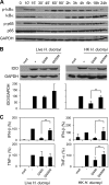
H. ducreyi-induced IDO expression in DC. IDO protein production by DC exposed to live (A) and heat-killed (B) H. ducreyi. Shown are representative Western blots of samples obtained from three donors. DC were incubated with medium (med) or with live or heat-killed H. ducreyi at MOI of 3:1 and 1:1, respectively, in the time course studies (left panels) and at various MOI for 24 h in the dose-response studies (right panels). (C) Induction of IDO enzymatic activity. DC were incubated with medium or with live or heat-killed H. ducreyi for 24 h, washed, and cultured in HBSS containing 100 μM tryptophan for 4 h. Supernatants were assessed for kynurenine production with the Ehrlich reagent. Bars represent the mean ± SD of results from six donors. *, P ≤ 0.05 compared with the medium-treated DC group (the Wilcoxon signed rank tests).
Fig. 2.
Fig. 2.
Critical role of type I interferons and TNF-α in H. ducreyi-induced IDO production. (A) DC were incubated with medium (med) and with live or heat-killed H. ducreyi in the presence of neutralizing antibodies to IFN-γ, IFNAR2, TNF-α, IFNAR2 and TNF-α, or isotype-matched control antibodies mIgG1 and mIgG2a. Top panels are representative Western blots, and bottom panels are densitometry data obtained from at least four individual donors. The ratio of IDO/GAPDH in each sample was normalized to that of DC incubated with H. ducreyi alone, which was set to a value of 100, and was expressed as a mean ± SD. *, P ≤ 0.05; **, P ≤ 0.01; ***, P ≤ 0.001. α, anti. (B) Time course of IFN-β and TNF-α production by DC stimulated with live or heat-killed H. ducreyi. Data represent results from multiple measurements made in one of two independent experiments.
Fig. 3.
Fig. 3.
Role of MAPK and PI3K activation in the expression of H. ducreyi-induced IDO, IFN-β, and TNF-α. (A) Time course of MAPK and PI3K activation. Western blots of whole-cell lysates of DC infected with live H. ducreyi for various times (in minutes) were probed with antibodies to phosphorylated or total forms of MAPKs and Akt. (B) Effects of MAPK and PI3K inhibitors on IDO expression. DC were incubated in medium (med) or infected with live H. ducreyi in the presence of SB203580 (SB; p38 inhibitor), SP600125 (SP; JNK inhibitor), U0126 (ERK inhibitor), the PI3K inhibitor wortmannin (WM), and the vehicle control DMSO. Top panels are representative Western blots, and the bottom panel shows densitometry data obtained from five donors. Bars represent mean ± SD of the ratio of IDO/GAPDH. (C) Effects of MAPK inhibitors on the production of IFN-β and TNF-α. Culture supernatants from DC infected for 6 and 24 h were assessed for the accumulation of IFN-β (n = 6) and TNF-α (n = 5), respectively. Due to donor-to-donor variation in cytokine production, the cytokine level in each sample was normalized to that of DC incubated with H. ducreyi alone, which was set at 100%. Bars represent mean ± SD. **, P ≤ 0.01; ***, P ≤ 0.001.
Fig. 4.
Fig. 4.
Role of NF-κB activation in the production of H. ducreyi-induced IDO, IFN-β, and TNF-α. (A) Time course of NF-κΒ activation. Western blots of whole-cell lysates of DC infected with live H. ducreyi for various periods of time were probed with antibodies against phosphorylated or total forms of IκBα and p65 and GAPDH. Shown are representative Western blots of samples obtained from three donors. (B) Effect of NF-κB inhibitor NF-κB SN50 on IDO production. DC were incubated in medium (med) or with live or heat-killed (HK) H. ducreyi in the presence or absence of NF-κB SN50 or its inactive control NF-κB SN50M. Top panels are representative Western blots, and bottom panels are densitometry data obtained from four donors. The ratio of IDO/GAPDH in each sample was normalized to that of DC incubated with H. ducreyi alone, which was set to a value of 100, and is expressed as a mean ± SD. (C) Effect of NF-κB SN50 on the production of IFN-β and TNF-α. In culture supernatants from DC infected with live H. ducreyi for 6 h and 24 h, the levels of IFN-β (n = 4) and TNF-α (n = 4), respectively, were measured. Supernatants from DC treated with heat-killed bacteria for 18 h were assessed for the production of IFN-β (n = 4) and TNF-α (n = 5). Due to donor-to-donor variation in cytokine production, cytokine levels were normalized to that of DC incubated with H. ducreyi alone, which was set at 100%. Bars are means ± SD. *, P ≤ 0.05; **, P ≤ 0.01.
Fig. 5.
Fig. 5.
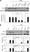
H. ducreyi LOS is a major inducer of IDO. (A) Nonessential role of bacterial ingestion in IDO production. DC were cultured in medium (med) or infected with live H. ducreyi in the presence of DMSO or cytochalasin D (CD) or with live H. ducreyi separated by a 0.2-μm-pore-size transwell (TW). Western blots represent results from five cytochalasin D and six transwell experiments, respectively. Bars represent mean ± SD of the ratio of IDO/GAPDH, which was determined by densitometry. (B) Inhibitory effect of polymyxin B (PMB) on the expression of IDO induced by H. ducreyi culture supernatants (sup) and purified H. ducreyi LOS. DC were stimulated with supernatants or LOS in the presence and 0, 10, or 30 μg/ml of polymyxin B. Shown are representative Western blots of samples obtained from five donors. (C) Effect of anti-TLR4 antibody and polymyxin B on H. ducreyi-induced IDO production. DC were stimulated with heat-killed bacteria in the presence of anti-TLR4 antibody or isotype-matched control antibody (iso) (left panels) or polymyxin B (right panels). Top panels are representative Western blots, and bottom panels are densitometry data obtained from four antibody inhibition and five polymyxin B inhibition experiments. The ratio of IDO/GAPDH in each sample was normalized to that of DC incubated with H. ducreyi alone, which was set to a value of 100, and is expressed as a mean ± SD. *, P ≤ 0.05. α, anti.
Fig. 6.
Fig. 6.
H. ducreyi LOS-induced IDO production is mediated by type I interferons, TNF-α, and the MAPK and NF-κB signaling pathways. (A) Effect of neutralizing antibodies against type I interferon receptor and TNF-α on IDO induction. DC were incubated in medium (med) or stimulated with LOS in the presence of antibodies against IFNAR2, TNF-α, IFNAR2 and TNF-α, or isotype-matched control antibodies mIgG1 and mIgG2a. Top panels are representative Western blots, and bottom panels are densitometry data obtained from four donors. The ratio of IDO/GAPDH in each sample was normalized to that of DC incubated with LOS alone, which was set to a value of 100, and is expressed as a mean ± SD. α, anti. (B) Inhibitory effect of the MAPK and NF-κΒ inhibitors on IDO induction. Cell lysates of DC treated with medium or LOS in the presence of DMSO, the MAPK inhibitors SB203580 (SB), SP600125 (SP), and U0126, or the NF-κB inhibitor NF-κB SN50 (SN50) and its inactive control NF-κB SN50M (SN50M) were analyzed for IDO and GAPDH protein expression. Representative Western blots of samples obtained from four donors are shown at the top. Culture supernatants from unstimulated DC or DC stimulated with LOS for 6 h were assayed for the production of IFN-β (n = 3) and TNF-α (n = 5 for the MAPK inhibitors and n = 3 for the NF-κB inhibitor), respectively. Due to donor-to-donor variation in cytokine production, cytokine levels were normalized to those of DC incubated with LOS alone, which was set at 100%. Bars indicate mean ± SD. *, P ≤ 0.05; **, P ≤ 0.01; ***, P ≤ 0.001.
Fig. 7.
Fig. 7.
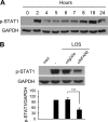
The JAK-STAT pathway is activated by H. ducreyi LOS-induced IFN-β. (A) Time course of STAT1 activation by LOS. Western blots of whole-cell lysates of DC stimulated with H. ducreyi LOS for various periods of time (hours) were probed with antibodies against phosphorylated STAT1 and GAPDH. Shown are representative Western blots of samples obtained from two donors. (B) Effect of neutralizing antibodies against type I interferon receptor on STAT1 activation. DC were incubated in medium (med) or stimulated with LOS in the presence of antibodies against IFNAR2 or isotype-matched control antibody mIgG2a. Top panels are representative Western blots, and bottom panels are densitometry data obtained from five donors. The ratio of phosphorylated STAT1/GAPDH in each sample was normalized to that of DC incubated with LOS alone, which was set to a value of 100, and is expressed as a mean ± SD. ***, P ≤ 0.001.
Fig. 8.
Fig. 8.
Schematic overview of IDO induction by H. ducreyi LOS. Engagement of TLR4 on the DC surface by H. ducreyi LOS activates MyD88-dependent and MyD88-independent signaling pathways. The MyD88-dependent pathway leads to the activation of MAPK (p38, JNK, and ERK) and NF-κB signals. The MyD88-independent pathway is mediated by TRIF, which activates IRF3 as well as MAPKs and NF-κB. Signaling through MAPKs and NF-κB leads to the expression of TNF-α, whereas activation of IRF3 (27, 46), MAPKs, and NF-κB is required for the expression of IFN-β. Secreted TNF-α and IFN-β bind to their cognate receptors, TNF receptor (TNFR) and IFNAR, via autocrine mechanisms to activate the MAPK and NF-κB and JAK-STAT signal pathways, respectively. The JAK-STAT signaling promotes the formation of the transcription factor complex ISGF3 (a heterotrimer of STAT1, STAT2, and IRF9). The cooperative activation of these pathways leads to transcriptional activation of the INDO gene, which encodes IDO, and the production of IDO protein.

Similar articles

Cited by

References

    1. Adams O., et al. 2004. Inhibition of human herpes simplex virus type 2 by interferon gamma and tumor necrosis factor alpha is mediated by indoleamine 2,3-dioxygenase. Microbes Infect. 6:806–812 - PubMed
    1. Baban B., et al. 2009. IDO activates regulatory T cells and blocks their conversion into Th17-like T cells. J. Immunol. 183:2475–2483 - PMC - PubMed
    1. Baban B., et al. 2005. A minor population of splenic dendritic cells expressing CD19 mediates IDO-dependent T cell suppression via type I IFN signaling following B7 ligation. Int. Immunol. 17:909–919 - PubMed
    1. Babcock T. A., Carlin J. M. 2000. Transcriptional activation of indoleamine dioxygenase by interleukin 1 and tumor necrosis factor alpha in interferon-treated epithelial cells. Cytokine 12:588–594 - PubMed
    1. Banks K. E., et al. 2008. The enterobacterial common antigen-like gene cluster of Haemophilus ducreyi contributes to virulence in humans. J. Infect. Dis. 197:1531–1536 - PubMed

Publication types

MeSH terms