. 2011 Jul;79(7):2829-38.
doi: 10.1128/IAI.00097-11. Epub 2011 May 16.

Spontaneous phthiocerol dimycocerosate-deficient variants of Mycobacterium tuberculosis are susceptible to gamma interferon-mediated immunity

Affiliations

Spontaneous phthiocerol dimycocerosate-deficient variants of Mycobacterium tuberculosis are susceptible to gamma interferon-mediated immunity

Meghan A Kirksey et al. Infect Immun. 2011 Jul.

Abstract

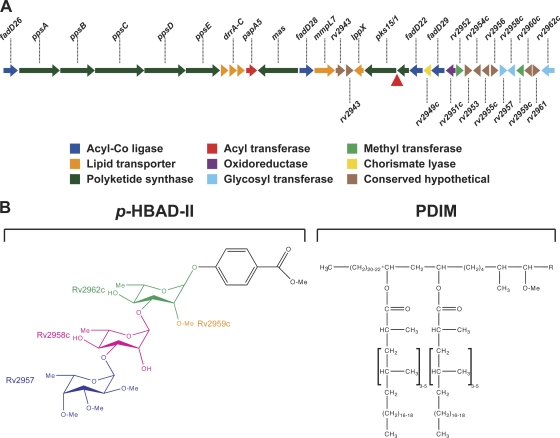
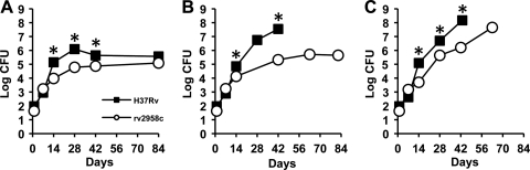
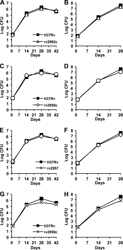
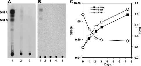
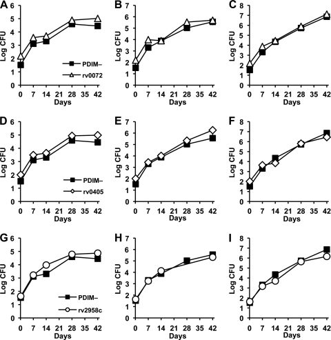
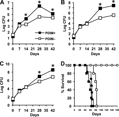
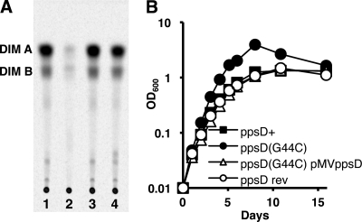
Onset of the adaptive immune response in mice infected with Mycobacterium tuberculosis is accompanied by slowing of bacterial replication and establishment of a chronic infection. Stabilization of bacterial numbers during the chronic phase of infection is dependent on the activity of the gamma interferon (IFN-γ)-inducible nitric oxide synthase (NOS2). Previously, we described a differential signature-tagged mutagenesis screen designed to identify M. tuberculosis "counterimmune" mechanisms and reported the isolation of three mutants in the H37Rv strain background containing transposon insertions in the rv0072, rv0405, and rv2958c genes. These mutants were impaired for replication and virulence in NOS2(-/-) mice but were growth-proficient and virulent in IFN-γ(-/-) mice, suggesting that the disrupted genes were required for bacterial resistance to an IFN-γ-dependent immune mechanism other than NOS2. Here, we report that the attenuation of these strains is attributable to an underlying transposon-independent deficiency in biosynthesis of phthiocerol dimycocerosate (PDIM), a cell wall lipid that is required for full virulence in mice. We performed whole-genome resequencing of a PDIM-deficient clone and identified a spontaneous point mutation in the putative polyketide synthase PpsD that results in a G44C amino acid substitution. We demonstrate by complementation with the wild-type ppsD gene and reversion of the ppsD gene to the wild-type sequence that the ppsD(G44C) point mutation is responsible for PDIM deficiency, virulence attenuation in NOS2(-/-) and wild-type C57BL/6 mice, and a growth advantage in vitro in liquid culture. We conclude that PDIM biosynthesis is required for M. tuberculosis resistance to an IFN-γ-mediated immune response that is independent of NOS2.


Figures

Fig. 1.
Fig. 1.
PDIM and p-HBAD biosynthesis in M. tuberculosis. (A) Genomic locus responsible for PDIM and p-HBAD-II biosynthesis. In M. tuberculosis H37Rv and other members of the Euro-American lineage, a frameshift mutation (red triangle) disrupts the pks15/1 open reading frame. Other M. tuberculosis lineages have an intact pks15/1 locus that encodes a functional polyketide synthase. (B) Structures of p-HBAD-II and PDIM. The polyketide synthase Pks15/1 adds malonyl coenzyme A (malonyl-CoA) units to p-hydroxybenzoic acid to generate p-hydroxyphenylalkanoic acid derivatives, which are precursors of PGL biosynthesis (5). M. tuberculosis 37Rv lacks Pks15/1 activity and produces tri-glycosylated p-hydroxybenzoic acid (p-HBAD-II) and PDIM instead. The Rv2962c, Rv2958c, and Rv2957 glycosyl transferases add rhamnose → rhamnose → fucose to p-hydroxyphenylalkanoic acid (25), and the Rv2959c methyltransferase O-methylates the C2 ring position of the proximal rhamnosyl residue (24).
Fig. 2.
Fig. 2.
Growth kinetics of M. tuberculosis strains H37Rv (wild type) and rv2958c::Tn in wild-type and immunodeficient mice. C57BL/6 (A), NOS2−/− (B), and IFN-γ−/− (C) mice were aerosol infected with M. tuberculosis strains H37Rv (squares) or rv2958c::Tn (circles). These strains were described previously (12). Groups of mice were sacrificed at the indicated time points, and bacterial CFU were enumerated by plating lung homogenates on 7H10 agar and scoring colonies after 3 to 4 weeks of incubation at 37°C. Symbols represent means (n = 4 or 5 mice per group per time point); error bars indicate standard errors. Asterisks indicate a statistically significant difference (P < 0.05) between the groups. Representative results of two independent experiments are shown.
Fig. 3.
Fig. 3.
p-HBAD-II biosynthesis is not required for M. tuberculosis growth and survival in wild-type or immunodeficient mice. C57BL/6 (A, C, E, and G) and NOS2−/− (B, D, F, and H) mice were aerosol infected with M. tuberculosis strains H37Rv (A to H, squares), Δrv2962c (A and B, triangles), Δrv2958c (C and D, circles), Δrv2958c (E and F, diamonds), or Δrv2959c (G and H, crosses). These strains were described previously (24, 25). Groups of mice were sacrificed at the indicated time points, and bacterial CFU were enumerated by plating lung homogenates on 7H10 agar and scoring colonies after 3 to 4 weeks of incubation at 37°C. Symbols represent means (n = 4 or 5 mice per group per time point); error bars indicate standard errors. This experiment was performed once.
Fig. 4.
Fig. 4.
PDIM deficiency confers an in vitro growth advantage in M. tuberculosis H37Rv. (A and B) Thin-layer chromatographic analysis of PDIM biosynthesis. Bacteria were labeled with [14C]propionate, which preferentially labels PDIM (6), and cell wall lipids were extracted and separated by thin-layer chromatography. (A) M. tuberculosis strains. H37Rv, Guilhot lab (25) (lane 1); H37Rv, McKinney lab (12) (lane 2); rv2958c::Tn (12) (lane 3). (B) M. tuberculosis strains, McKinney lab (12). H37Rv (lane 1), H37Rv after subculture (lane 2), rv2958c::Tn (lane 3), rv0072::Tn (lane 4), rv0405::Tn (lane 5). (C) Independently derived subclones of PDIM-positive H37Rv (squares) and PDIM-negative H37Rv (circles) were grown in 7H9 broth with aeration at 37°C. Growth of the cultures was monitored by withdrawing aliquots and measuring the OD600 at the indicated time points (plotted on the primary y axis). The (PDIM-positive OD600)/(PDIM-negative OD600) ratios at each time point are plotted on the secondary y axis (diamonds). Results are representative of three independent experiments.
Fig. 5.
Fig. 5.
Growth kinetics of PDIM-negative H37Rv and M. tuberculosis rv0072::Tn, rv0405::Tn, and rv2958c::Tn mutants in wild-type and immunodeficient mice. C57BL/6 (A, D, and G), NOS2−/− (B, E, and H), and IFN-γ−/− (C, F, and I) mice were aerosol infected with M. tuberculosis H37Rv PDIM-negative variant (A to I, squares), rv0072::Tn (A to C, triangles), rv0405::Tn (D to F, diamonds), or rv2958c::Tn (G to I, circles). The PDIM-negative variant of H37Rv is described herein; the Tn mutant strains were described previously (12). Groups of mice were sacrificed at the indicated time points, and bacterial CFU were enumerated by plating lung homogenates on 7H10 agar and scoring colonies after 3 to 4 weeks of incubation at 37°C. Symbols represent means (n = 4 or 5 mice per group per time point); error bars indicate standard errors. This experiment was performed once.
Fig. 6.
Fig. 6.
Growth kinetics of PDIM-positive and PDIM-negative subclones of M. tuberculosis H37Rv in wild-type and immunodeficient mice. (A to C) C57BL/6 (A), NOS2−/− (B), and IFN-γ−/− (C) mice were aerosol-infected with PDIM-positive (filled squares) or PDIM-negative (open squares) subclones of M. tuberculosis H37Rv. Groups of mice were sacrificed at the indicated time points, and bacterial CFU were enumerated by plating lung homogenates on 7H10 agar and scoring colonies after 3 to 4 weeks of incubation at 37°C. Symbols represent means (n = 4 or 5 mice per group per time point); error bars indicate standard errors. Asterisks indicate statistically significant differences (P < 0.05) between the groups. (D) Survival of NOS2−/− (circles) and IFN-γ−/− (squares) mice (n = 5 or 6 per group) after aerosol infection with PDIM-positive (filled symbols) or PDIM-negative (open symbols) subclones of M. tuberculosis H37Rv. This experiment was performed once.
Fig. 7.
Fig. 7.
The ppsD(G44C) point mutation is responsible for PDIM deficiency and the in vitro growth advantage of the H37Rv PDIM-negative subclone. (A) Thin-layer chromatographic analysis of PDIM biosynthesis. Bacteria were labeled with [14C]propionate, which is preferentially incorporated into PDIM (6), and cell wall lipids were extracted and separated by thin-layer chromatography. M. tuberculosis H37Rv strains: ppsD+ (lane 1), ppsD(G44C) (lane 2), ppsD rev (lane 3), ppsD(G44C) pMV361-ppsD (lane 4). Results are representative of two independent experiments. (B) Bacteria were grown in 7H9 broth with aeration at 37°C. Growth was monitored by withdrawing aliquots and measuring the OD600 at the indicated time points. M. tuberculosis H37Rv strains: ppsD+ (filled squares), ppsD(G44C) (filled circles), ppsD rev (open circles), ppsD(G44C) pMV361-ppsD (open triangles). Results are representative of three independent experiments.
Fig. 8.
Fig. 8.
Reversion of the ppsD(G44C) point mutation restores wild-type levels of growth and virulence in mice. (A to D) Mice were aerosol infected with ppsD+ (filled squares), ppsD(G44C) (filled circles), or ppsD rev (open circles) strains of M. tuberculosis H37Rv. (A and B) Bacterial growth in the lungs of C57BL/6 (A) and NOS2−/− (B) mice. Groups of mice were sacrificed at the indicated time points, and bacterial CFU were enumerated by plating lung homogenates on 7H10 and scoring colonies after 3 to 4 weeks of incubation at 37°C. Symbols represent means (n = 4 mice per group); error bars indicate standard errors. Asterisks indicate statistically significant differences (P < 0.05) in comparisons of ppsD(G44C) versus ppsD+ and ppsD(G44C) versus ppsD rev strains. This experiment was performed once. (C and D) Survival of IFN-γ−/− (C) and iNOS−/− (D) mice (n = 5 mice per group). This experiment was performed once.

References

    1. Andreu N., Gibert I. 2008. Cell population heterogeneity in Mycobacterium tuberculosis H37Rv. Tuberculosis (Edinb.) 88:553–559 - PubMed
    1. Astarie-Dequeker C., et al. 2009. Phthiocerol dimycocerosates of M. tuberculosis participate in macrophage invasion by inducing changes in the organization of plasma membrane lipids. PLoS Pathog. 5:e1000289. - PMC - PubMed
    1. Camacho L. R., et al. 2001. Analysis of the phthiocerol dimycocerosate locus of Mycobacterium tuberculosis: evidence that this lipid is involved in the cell wall permeability barrier. J. Biol. Chem. 276:19845–19854 - PubMed
    1. Camacho L. R., Ensergueix D., Perez E., Gicquel B., Guilhot C. 1999. Identification of a virulence gene cluster of Mycobacterium tuberculosis by signature-tagged transposon mutagenesis. Mol. Microbiol. 34:257–267 - PubMed
    1. Constant P., et al. 2002. Role of the pks15/1 gene in the biosynthesis of phenolglycolipids in the Mycobacterium tuberculosis complex: evidence that all strains synthesize glycosylated p-hydroxybenzoic acid methyl esters and that strains devoid of phenolglycolipids harbor a frameshift mutation in the pks15/1 gene. J. Biol. Chem. 277:38148–38158 - PubMed

Publication types

MeSH terms

LinkOut - more resources