. 2011 Jun 6;208(6):1243-52.
doi: 10.1084/jem.20102477. Epub 2011 May 16.

A dynamic T cell-limited checkpoint regulates affinity-dependent B cell entry into the germinal center

Affiliations

A dynamic T cell-limited checkpoint regulates affinity-dependent B cell entry into the germinal center

Tanja A Schwickert et al. J Exp Med. .

Abstract

The germinal center (GC) reaction is essential for the generation of the somatically hypermutated, high-affinity antibodies that mediate adaptive immunity. Entry into the GC is limited to a small number of B cell clones; however, the process by which this limited number of clones is selected is unclear. In this study, we demonstrate that low-affinity B cells intrinsically capable of seeding a GC reaction fail to expand and become activated in the presence of higher-affinity B cells even before GC coalescence. Live multiphoton imaging shows that selection is based on the amount of peptide-major histocompatibility complex (pMHC) presented to cognate T cells within clusters of responding B and T cells at the T-B border. We propose a model in which T cell help is restricted to the B cells with the highest amounts of pMHC, thus allowing for a dynamic affinity threshold to be imposed on antigen-binding B cells.


Figures

Figure 1.
Figure 1.
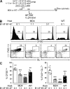
GC occupancy is regulated by competition. (A) Diagrammatic representation of the experimental protocol. (B) Histology showing composition of GCs in draining lymph nodes 6 d after immunization. B1-8lo B cells are GFP+ (false-colored red), and B1-8hi B cells are CD45.1+ (green). GCs were identified by absence of IgD+ B cells (blue) within the B cell follicle. Images are representative of multiple GCs from two independent experiments. (C) Composition of GCs by flow cytometry. Gated on CD19+Igλ+FAS+GL-7+. (left) Representative flow cytometry plots showing the fraction of cells of each genotype (B1-8lo B cells are CFP+, B1-8hi B cells are CD45.1+, and endogenous B cells are CFPCD45.1). (right) Quantification of GC occupancy under different conditions. Each symbol represents one mouse. Data are from two independent experiments with two to three mice per condition per experiment.
Figure 2.
Figure 2.
Competition inhibits early proliferation and activation of low-affinity B cells. (A) Diagrammatic representation of the experimental protocol. (B) Flow cytometric analysis of transferred B1-8lo CD45.1+ CFSE-labeled B cells in draining lymph nodes 4 d after immunization. Gated on CD19+Igλ+CD45.1+ cells. (top) Representative CFSE dilution histograms. (bottom) Representative FAS/GL-7 dot plots. (C) Quantification of B. (left) Percentage of divided (CFSElow) B1-8lo B cells. (right) Percentage of FASintGL-7+ B1-8lo cells. Each symbol represents one mouse. Error bars are SEM. Data are from three independent experiments with one to three mice per condition per experiment. **, P < 0.01; ***, P < 0.001 compared with B1-8lo in MD4 host.
Figure 3.
Figure 3.
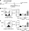
Competition does not affect antigen binding and presentation by low-affinity B cells. (A) Diagrammatic representation of the experimental protocol. (B, left) Flow cytometric analysis of antigen binding by B1-8hi and B1-8lo cells under different transfer conditions. Gated as CD19+Igλ+CD45.1+ (B1-8hi) or CD19+Igλ+CFP+ (B1-8lo). (right) Quantification of flow cytometry data. (C, left) Flow cytometric analysis of antigen presentation by B1-8hi and B1-8lo cells under different transfer conditions. Cell types are gated as in B; only Alexa Fluor 647+ cells are plotted. The Y-Ae antibody binds to the I-Eα52–68 peptide in the context of I-Ab. (right) Quantification of flow cytometry data. Each symbol represents one mouse. Data are from two independent experiments with two mice per condition per experiment.
Figure 4.
Figure 4.
Inhibition of proliferation/activation by competition is independent of antigen dose. (A) Diagrammatic representation of the experimental protocol. (B) Flow cytometric analysis of transferred B1-8lo CD45.1+ CFSE-labeled B cells in draining lymph nodes 4 d after immunization. Gated on CD19+Igλ+CD45.1+. (top) Representative FAS/GL-7 plots. (bottom) Representative CFSE histogram. (C) Quantification of data in B. Each symbol represents one mouse. Data are from two independent experiments with two to three mice per condition per experiment.
Figure 5.
Figure 5.
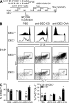
Access to T cell help drives early B cell competition. (A) Diagrammatic representation of the experimental protocol. (B) Flow cytometric analysis of transferred B1-8hiDEC205+/+CFP+ and B1-8hiDEC205−/−CD45.1+ CFSE-labeled B cells in draining lymph nodes 4 d after immunization. Gated on CD19+Igλ+CD45.1+ (B1-8hiDEC205+/+) or CD19+Igλ+CFP+ (B1-8hiDEC205+/+) cells. (top) Representative CFSE dilution profile. (bottom) Representative FAS/GL-7 plot. (C) Quantification of data in B. Each symbol represents one mouse. Data are from three independent experiments with one to two mice per condition per experiment. CS, P. falciparum circumsporozoite protein (used as a control antigen).
Figure 6.
Figure 6.
Increased T cell help drives greater GC occupancy and plasmablast differentiation. (A) Diagrammatic representation of the experimental protocol. (B) Flow cytometric analysis of transferred B1-8hiDEC205+/+CFP+ and B1-8hiDEC205−/−CD45.1+ B cells in draining lymph nodes 6 d after immunization. (top) GC occupancy. Gated on CD19+Igλ+GL-7+FAS+. (bottom) Composition of plasmablast compartment. Gated on CD138hiB220int. (C) Quantification of data in B. Each symbol represents one mouse. Data are from three independent experiments with one to two mice per condition per experiment. CS, P. falciparum circumsporozoite protein (used as a control antigen).
Figure 7.
Figure 7.
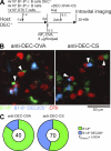
B cells with higher surface MHC preferentially interact with cognate T cells at the T–B border. (A) Diagrammatic representation of the experimental protocol. (B) Collapsed z stacks of three-dimensional image series of T–B border clusters in living mice. Arrowheads indicate long-lasting conjugates (>10 min) between OT-II T cells (red) and either B1-8hiDEC205+/+ (green) or B1-8hiDEC205−/− (cyan) B cells. (C) Quantification of T–B cell interactions in intravital videos. Pie charts represent the proportion of long-lasting T–B conjugates involving DEC205+/+ and DEC205−/− B1-8hi B cells. The total number of conjugates analyzed is indicated in the center of each chart. The charts represent pooled data from multiple videos taken from three independent experiments, one mouse per condition per experiment. CS, P. falciparum circumsporozoite protein (used as a control antigen).

References

    1. Allen C.D., Okada T., Tang H.L., Cyster J.G. 2007. Imaging of germinal center selection events during affinity maturation. Science. 315:528–531 10.1126/science.1136736 - DOI - PubMed
    1. Allen D., Simon T., Sablitzky F., Rajewsky K., Cumano A. 1988. Antibody engineering for the analysis of affinity maturation of an anti-hapten response. EMBO J. 7:1995–2001 - PMC - PubMed
    1. Barnden M.J., Allison J., Heath W.R., Carbone F.R. 1998. Defective TCR expression in transgenic mice constructed using cDNA-based alpha- and beta-chain genes under the control of heterologous regulatory elements. Immunol. Cell Biol. 76:34–40 10.1046/j.1440-1711.1998.00709.x - DOI - PubMed
    1. Bonifaz L., Bonnyay D., Mahnke K., Rivera M., Nussenzweig M.C., Steinman R.M. 2002. Efficient targeting of protein antigen to the dendritic cell receptor DEC-205 in the steady state leads to antigen presentation on major histocompatibility complex class I products and peripheral CD8+ T cell tolerance. J. Exp. Med. 196:1627–1638 10.1084/jem.20021598 - DOI - PMC - PubMed
    1. Boscardin S.B., Hafalla J.C., Masilamani R.F., Kamphorst A.O., Zebroski H.A., Rai U., Morrot A., Zavala F., Steinman R.M., Nussenzweig R.S., Nussenzweig M.C. 2006. Antigen targeting to dendritic cells elicits long-lived T cell help for antibody responses. J. Exp. Med. 203:599–606 10.1084/jem.20051639 - DOI - PMC - PubMed

Publication types