Review
. 2011 May 16;193(4):607-18.
doi: 10.1083/jcb.201012094.

Lymphatic vascular morphogenesis in development, physiology, and disease

Affiliations
Review

Lymphatic vascular morphogenesis in development, physiology, and disease

Stefan Schulte-Merker et al. J Cell Biol. .

Abstract

The lymphatic vasculature constitutes a highly specialized part of the vascular system that is essential for the maintenance of interstitial fluid balance, uptake of dietary fat, and immune response. Recently, there has been an increased awareness of the importance of lymphatic vessels in many common pathological conditions, such as tumor cell dissemination and chronic inflammation. Studies of embryonic development and genetically engineered animal models coupled with the discovery of mutations underlying human lymphedema syndromes have contributed to our understanding of mechanisms regulating normal and pathological lymphatic morphogenesis. It is now crucial to use this knowledge for the development of novel therapies for human diseases.


Figures
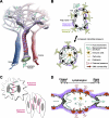
Figure 1.
Figure 1.
Organization of lymphatic vasculature. (A) The lymphatic vasculature resorbs fluid, macromolecules, and cells from the interstitium. (B) Mechanism of lymph formation in capillaries. Interstitial components penetrate lymphatic capillaries via openings between LECs. The specialized structure of such openings prevents the return of lymph back to the interstitium. Anchoring filaments attach LECs to the ECM and prevent vessel collapse under conditions of increased interstitial pressure (black arrow). (C) Junctional organization of LECs in lymphatic capillaries and collecting vessels. Both “buttons” and “zippers” share a repertoire of adherens and tight junction–associated proteins (e.g., VE-cadherin, zonula occludens-1, occludin, and claudin-5). The main difference between them resides in their organization (Baluk et al., 2007). (D) Mechanism of lymph propulsion in collecting vessels. Coordinated opening and closure of lymphatic valves is important for efficient lymph transport. SMCs covering each lymphangion possess intrinsic contractile activity. EC, endothelial cell.
Figure 2.
Figure 2.
Main steps of mammalian lymphatic vascular development. (A) LECs are specified in embryonic veins, from where they sprout toward Vegf-c–producing mesodermal cells and aggregate to form lymph sacs. Further sprouting produces the lymphatic primary plexus composed of capillary-like vessels. Myeloid cells produce cytokines and regulate lymphatic vascular morphogenesis. Primary plexus is further remodeled to form collecting, precollecting, and capillary compartments. Precollecting and collecting lymphatic vessels have intraluminal valves and basement membrane coverage. Collecting lymphatic vessels are surrounded by SMCs (red). (box) Genes important for collecting lymphatic vessel development. (B) LYVE-1 is the earliest known LEC marker. The transcription factor Prox1 is essential for the establishment of LEC identity, and its expression is controlled by Sox18. (C) Signaling via Vegf-c and Vegfr-3 regulates LEC sprouting and proliferation. The role of Vegfr-2–Vegfr-3 heterodimers and participation of Nrp2 in the Vegfr-2–Vegfr-3 complex are not fully understood. (D) Separation of lymphatic and blood vasculature requires platelet aggregation (also see Table I). Interaction of podoplanin on LECs and CLEC-2 on platelets triggers the Syk-, Slp76-, and PLC-γ2–dependent signaling cascade leading to platelet aggregation. O-glycosylation by T-synthase is important for podoplanin function.
Figure 3.
Figure 3.
Causes of human hereditary lymphedemas. Lymph transport can be impaired because of a hypoplastic initial lymphatic capillary network, because of abnormal coverage of lymphatic capillaries with basement membrane components and SMCs or because of a lack of or malfunctioning lymphatic valves. Defective lymphatic drainage leads to tissue fibrosis and fat deposition caused by the abnormal local chronic inflammatory response. Genes that are mutated in human hereditary lymphedema are indicated in blue next to the processes to which they are thought to be causally related. Mechanisms of the action of GJC2, PTPN14, and IKBKG are not fully understood.

References

    1. Abtahian F., Guerriero A., Sebzda E., Lu M.M., Zhou R., Mocsai A., Myers E.E., Huang B., Jackson D.G., Ferrari V.A., et al. 2003. Regulation of blood and lymphatic vascular separation by signaling proteins SLP-76 and Syk. Science. 299:247–251 10.1126/science.1079477 - DOI - PMC - PubMed
    1. Adams R.H., Eichmann A. 2010. Axon guidance molecules in vascular patterning. Cold Spring Harb. Perspect. Biol. 2:a001875 10.1101/cshperspect.a001875 - DOI - PMC - PubMed
    1. Alders M., Hogan B.M., Gjini E., Salehi F., Al-Gazali L., Hennekam E.A., Holmberg E.E., Mannens M.M., Mulder M.F., Offerhaus G.J., et al. 2009. Mutations in CCBE1 cause generalized lymph vessel dysplasia in humans. Nat. Genet. 41:1272–1274 10.1038/ng.484 - DOI - PubMed
    1. Au A.C., Hernandez P.A., Lieber E., Nadroo A.M., Shen Y.M., Kelley K.A., Gelb B.D., Diaz G.A. 2010. Protein tyrosine phosphatase PTPN14 is a regulator of lymphatic function and choanal development in humans. Am. J. Hum. Genet. 87:436–444 10.1016/j.ajhg.2010.08.008 - DOI - PMC - PubMed
    1. Ayadi A., Zheng H., Sobieszczuk P., Buchwalter G., Moerman P., Alitalo K., Wasylyk B. 2001. Net-targeted mutant mice develop a vascular phenotype and up-regulate egr-1. EMBO J. 20:5139–5152 10.1093/emboj/20.18.5139 - DOI - PMC - PubMed

Publication types