Comment
. 2011 May 16;193(4):677-94.
doi: 10.1083/jcb.201012077.

Drosophila histone locus bodies form by hierarchical recruitment of components

Affiliations
Comment

Drosophila histone locus bodies form by hierarchical recruitment of components

Anne E White et al. J Cell Biol. .

Abstract

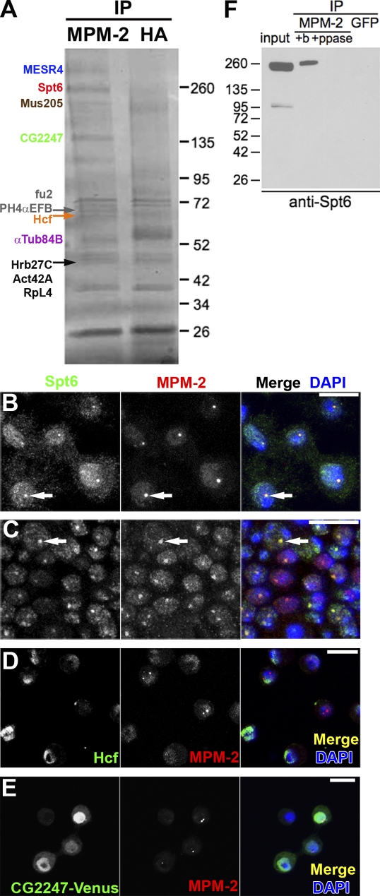
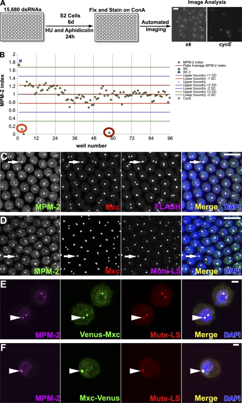
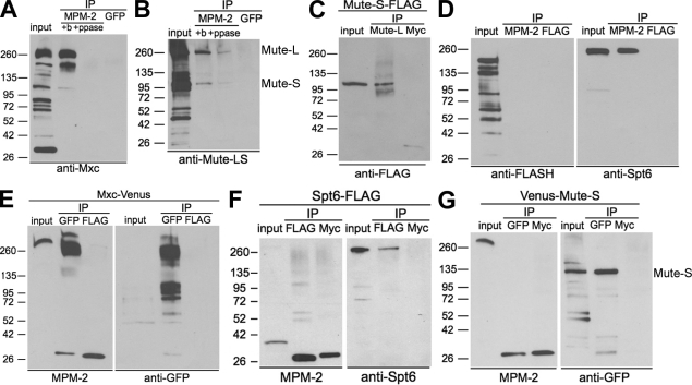
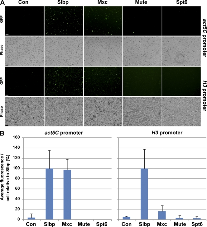
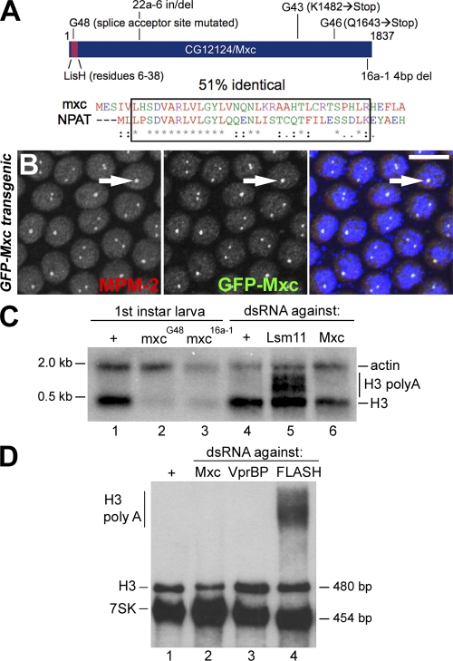
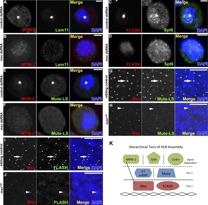
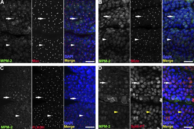
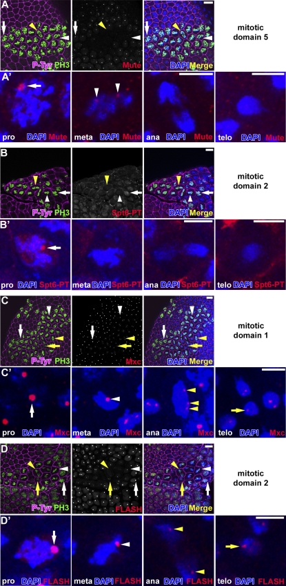
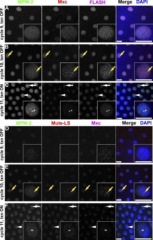
Nuclear bodies are protein- and RNA-containing structures that participate in a wide range of processes critical to genome function. Molecular self-organization is thought to drive nuclear body formation, but whether this occurs stochastically or via an ordered, hierarchical process is not fully understood. We addressed this question using RNAi and proteomic approaches in Drosophila melanogaster to identify and characterize novel components of the histone locus body (HLB), a nuclear body involved in the expression of replication-dependent histone genes. We identified the transcription elongation factor suppressor of Ty 6 (Spt6) and a homologue of mammalian nuclear protein of the ataxia telangiectasia-mutated locus that is encoded by the homeotic gene multisex combs (mxc) as novel HLB components. By combining genetic manipulation in both cell culture and embryos with cytological observations of Mxc, Spt6, and the known HLB components, FLICE-associated huge protein, Mute, U7 small nuclear ribonucleoprotein, and MPM-2 phosphoepitope, we demonstrated sequential recruitment and hierarchical dependency for localization of factors to HLBs during development, suggesting that ordered assembly can play a role in nuclear body formation.


Figures

Figure 1.
Figure 1.
Mass spectrometry identifies Spt6 as an HLB component. (A) Proteins precipitated with MPM-2 or control α-HA antibodies from S phase–arrested S2 cell nuclear extracts were separated by SDS-PAGE and stained with Coomassie. (B and C) S2 cells (B) and w1118 postblastoderm embryo (C) stained with α-Spt6, MPM-2, and DAPI. Spt6 forms nuclear foci that colocalize with MPM-2 (arrows). (D) S2 cells stained with α-Hcf, MPM-2, and DAPI. (E) S2 cells transiently transfected with CG2247-Venus and stained with α-GFP (green), MPM-2, and DAPI. Bars, 10 µm. (F) Nuclear extracts from S phase–arrested S2 cells were pretreated with buffer (+b) or λ phosphatase (+ppase) before immunoprecipitation (IP) with MPM-2 or control α-GFP antibodies and analyzed by Western blotting with α-Spt6. Molecular masses are given in kilodaltons.
Figure 2.
Figure 2.
Genome-wide RNAi screen for MPM-2–negative HLBs. (A) Overview of the screen. Bar, 5 µm. (B) Graph of the MPM-2 indices from a representative screen plate. Note the mean MPM-2 index of the plate (cyan +), three standard deviations from this mean (green lines), the cyclin E (CycE)–positive control (orange circle), and a hit (red circle). (C and D) w1118 syncytial blastoderm (cycle 13) embryos stained with MPM-2, α-Mxc, DAPI, and α-FLASH or α–Mute-LS. Arrows indicate HLBs. Bars, 10 µm. (E and F) S2 cells expressing Venus-Mxc or Mxc-Venus with the Actin5C promoter stained with MPM-2 and α-GFP (green), which recognizes Venus, α–Mute-LS, and DAPI. Arrowheads indicate HLBs. Two HLB foci arise from unpaired homologous chromosomes. Bars, 2 µm. ConA, concanavalin A; HU, hydroxyurea; SK, pBluescript.
Figure 3.
Figure 3.
Mxc is directly recognized by MPM-2. (A and B) Nuclear extracts from S phase–arrested S2 cells were pretreated with buffer only (+b) or λ phosphatase (+ppase) before immunoprecipitation (IP) with MPM-2 or GFP control antibodies and analyzed by Western blotting with α-Mxc (A) or α–Mute-LS (detects both the long and short isoforms of Mute; B). (C) α–Mute-L and α-Myc control IPs from cells expressing Mute-S–FLAG blotted with α-FLAG. (D) MPM-2 and α-FLAG control IPs from an S-phase–arrested S2 cell nuclear extract blotted with α-FLASH or α-Spt6. (E) α-GFP and α-FLAG control IPs from cells expressing Mxc-Venus blotted with MPM-2 or α-GFP. (F) α-FLAG and α-Myc control IPs from cells expressing Spt6-FLAG were blotted with α-Spt6 or MPM-2. (G) α-GFP and α-Myc control IPs from cells expressing Venus–Mute-S blotted with MPM-2 or α-GFP. Molecular masses are given in kilodaltons.
Figure 4.
Figure 4.
GFP histone mRNA misprocessing reporter analysis. Dmel-2 cells stably transfected with the indicated GFP histone mRNA misprocessing reporter were treated for 5 d with dsRNAs directed against control (Con; PTB), SLBP, Mxc, Mute, or Spt6 dsRNAs. (A) Representative epifluorescence and bright-field images. Bars, 60 µm. (B) Quantification of GFP fluorescence/cell for each experiment. SLBP was set as 100 for each reporter. Error bars indicate standard deviations. Between 200 and 800 cells were quantified per experiment.
Figure 5.
Figure 5.
Mxc is essential for histone gene expression. (A, top) Diagram of CG12124/Mxc showing the position of the LisH motif and mutations G48 (AG to AA at the first intron/second exon border), 22a-6 (GTGTCAGCTG insertion for AA at N480), G43 (K1482→Stop), G46 (Q1643→Stop), and 16a-1 (TTCG 4-bp frame-shifting deletion [4bp del] at F1823 changing 5′-GAGTTCGAGGACATC-3′ to 5′-GAGAGGACATC-3′). (bottom) ClustalW alignment of the LisH domain from Mxc and NPAT. Red, green, blue, and purple letters indicate hydrophobic, hydrophilic, acidic, and basic amino acids, respectively. The box indicates the LisH domain. The asterisks and dots indicate identical and conserved amino acids, respectively. Single dots are used to show positions of similarity; double dots are used to show positions of high conservation. (B) Syncytial blastoderm (cycle 12) embryos expressing GFP-Mxc stained with α-GFP, MPM-2, and DAPI (blue). Bar, 10 µm. (C and D) H3 Northern analysis of the indicated genotypes or 7-d dsRNA treatments. A plus sign indicates wild-type larva (C, lane 1) or mock RNAi (C and D, lane 1). VprBP is a nonspecific control. Actin (C) and 7SK (D) were used as loading controls. There is 1.5 times as much RNA loaded in D, lane 2 than lanes 1, 3, and 4.
Figure 6.
Figure 6.
HLB formation is disrupted in Mxc-depleted cells. (A–F) S2 cells treated with control (A, C, and E) or mxc (B, D, and F) dsRNAs were stained with MPM-2 (A, B, E, and F), α-Lsm11 (A and B), α-Spt6 (C and D), or α–Mute-LS (E and F) and DAPI. Bars, 2 µm. Note that DAPI staining is not uniform, and under the conditions of imaging the entire nucleus is not always visible. (G–J) Sibling control (G and I) and mxcG48 mutant (H and J) embryos stained with α-Mxc, DAPI, and α–Mute-LS (G and H) or α-FLASH (I and J). Arrows indicate HLBs, and arrowheads indicate an incomplete HLB. Bars, 10 µm. (K) Hierarchical model of Drosophila HLB assembly. Tier 1 and tier 2 are independent of histone gene expression, and HLB localization of tier 2 factors depends on tier 1 factors. Components above the horizontal dotted line are recruited to the HLB in response to particular signals: e.g., transcription for Spt6 and cyclin E/Cdk2 activity for the MPM-2 epitope. Signals that recruit coilin to the HLB are not known. The vertical dotted lines indicate no known interdependency between these factors for HLB localization. The line connecting MPM-2 and Mxc indicates that MPM-2 antibodies bind Mxc.
Figure 7.
Figure 7.
Spt6, but not Mxc, FLASH, or Mute, localization to HLBs is cell cycle dependent. (A–C) Stage 12 w1118 embryos were stained with MPM-2, DAPI, and α-Mxc (A), α–Mute-LS (B), or α-FLASH (C). (D) An embryo similarly stained using α-GFP to detect the Spt6 protein trap fusion protein. Arrows indicate S-phase cells containing MPM-2 foci that colocalize with Mxc (A), Mute-LS (B), FLASH (C), and Spt6-EGFP (D). White arrowheads indicate G117 cells lacking MPM-2 foci but containing Mxc (A), Mute-LS (B), and FLASH (C) in HLBs. Yellow arrowheads indicate a G117 cell lacking foci of MPM-2 and Spt6-EGFP. Thoracic segments 1 and 2 are shown with anterior at the top and ventral on the left. Bars, 10 µm.
Figure 8.
Figure 8.
Spt6 localizes to HLBs when histone genes are transcribed. (A) Postblastoderm Spt6-EGFP embryo entering mitosis in cell cycle 14 stained with MPM-2, α-GFP (green), and DAPI. Prominent Spt6 foci form in late G2 cells (arrows) but not in early G2 cells (arrowheads). Single late and early G2 cells are shown in B and C, respectively. Bars, 10 µm.
Figure 9.
Figure 9.
Mxc and FLASH remain chromosome associated during mitosis. (A–D) Postblastoderm w1118 embryos in cell cycle 14 stained with α–P-Tyr, α-PH3 (marks condensed, mitotic chromatin), DAPI, and α-Mute-LS (A), α-GFP (B), α-Mxc (C), or α-FLASH (D). Prophase (pro; white arrows), metaphase (meta; white arrowheads), anaphase (ana; yellow arrowheads), and telophase (telo; yellow arrows) cells are indicated. Bars, 10 µm. (A′–D′) Single prophase, metaphase, anaphase, and telophase cells are shown in A′, B′, C′, and D′. Bars, 5 µm. Note that the mouse α-PH3 used in D stains interphase cells weakly.
Figure 10.
Figure 10.
HLB assembly begins during nuclear cycle 10. (A–C) w1118 syncytial blastoderm embryos stained with MPM-2 (left), α-Mxc (middle), α-FLASH (right), and DAPI. (D–F) w1118 syncytial blastoderm embryos stained with MPM-2 (left), α–Mute-LS (middle), α-Mxc (right), and DAPI. Insets show a single nucleus. Interphase of nuclear cycles 9–11 is shown. Yellow arrows indicate a nucleus containing foci of Mxc and FLASH (B) or Mute (E) lacking a MPM-2 focus. Nuclei with one (white arrows) or two (white arrowheads) HLBs are indicated. txn, histone transcription. Bars, 10 µm.

Comment in

Comment on

References

    1. Andrulis E.D., Guzmán E., Döring P., Werner J., Lis J.T. 2000. High-resolution localization of Drosophila Spt5 and Spt6 at heat shock genes in vivo: roles in promoter proximal pausing and transcription elongation. Genes Dev. 14:2635–2649 10.1101/gad.844200 - DOI - PMC - PubMed
    1. Andrulis E.D., Werner J., Nazarian A., Erdjument-Bromage H., Tempst P., Lis J.T. 2002. The RNA processing exosome is linked to elongating RNA polymerase II in Drosophila. Nature. 420:837–841 10.1038/nature01181 - DOI - PubMed
    1. Ardehali M.B., Yao J., Adelman K., Fuda N.J., Petesch S.J., Webb W.W., Lis J.T. 2009. Spt6 enhances the elongation rate of RNA polymerase II in vivo. EMBO J. 28:1067–1077 10.1038/emboj.2009.56 - DOI - PMC - PubMed
    1. Barcaroli D., Bongiorno-Borbone L., Terrinoni A., Hofmann T.G., Rossi M., Knight R.A., Matera A.G., Melino G., De Laurenzi V. 2006a. FLASH is required for histone transcription and S-phase progression. Proc. Natl. Acad. Sci. USA. 103:14808–14812 10.1073/pnas.0604227103 - DOI - PMC - PubMed
    1. Barcaroli D., Dinsdale D., Neale M.H., Bongiorno-Borbone L., Ranalli M., Munarriz E., Sayan A.E., McWilliam J.M., Smith T.M., Fava E., et al. 2006b. FLASH is an essential component of Cajal bodies. Proc. Natl. Acad. Sci. USA. 103:14802–14807 10.1073/pnas.0604225103 - DOI - PMC - PubMed

Publication types

MeSH terms