. 2011 May 16;193(4):727-39.
doi: 10.1083/jcb.201101109.

The conversion of centrioles to centrosomes: essential coupling of duplication with segregation

Affiliations

The conversion of centrioles to centrosomes: essential coupling of duplication with segregation

Won-Jing Wang et al. J Cell Biol. .

Abstract

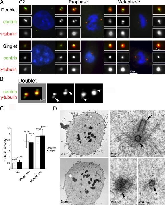
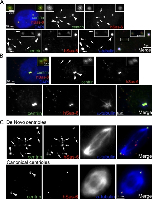
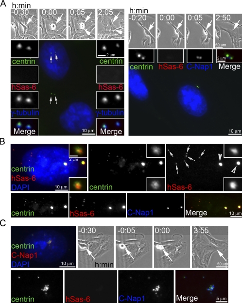
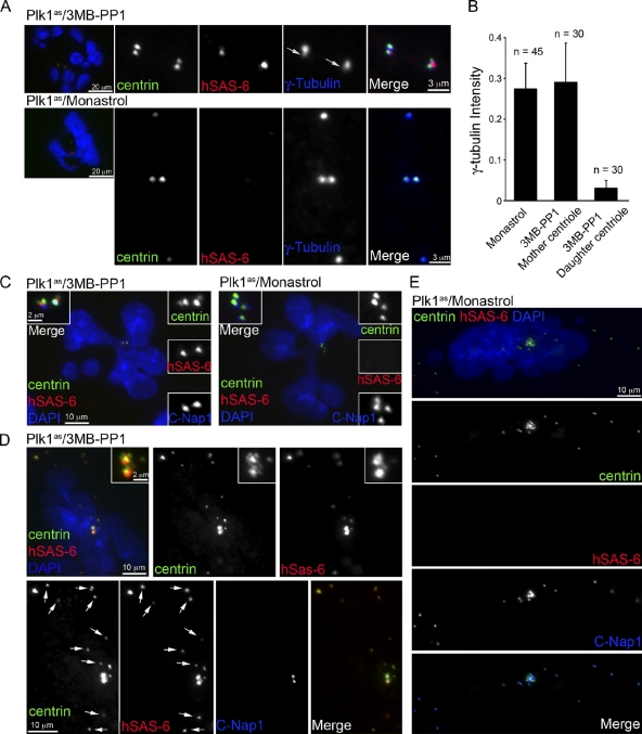
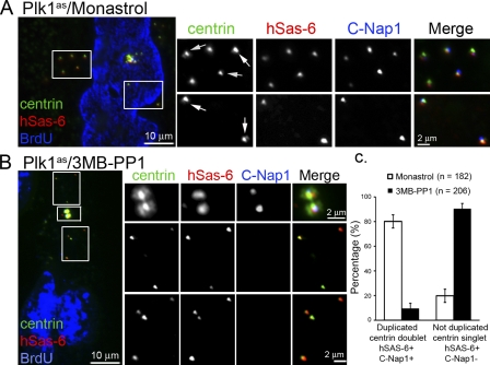
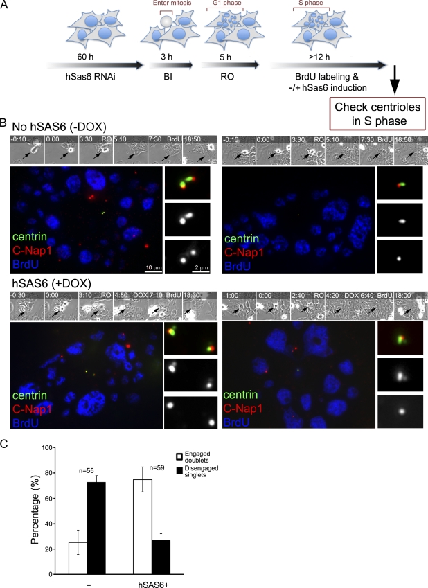
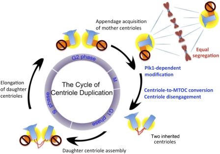
Centrioles are self-reproducing organelles that form the core structure of centrosomes or microtubule-organizing centers (MTOCs). However, whether duplication and MTOC organization reflect innate activities of centrioles or activities acquired conditionally is unclear. In this paper, we show that newly formed full-length centrioles had no inherent capacity to duplicate or to organize pericentriolar material (PCM) but acquired both after mitosis through a Plk1-dependent modification that occurred in early mitosis. Modified centrioles initiated PCM recruitment in G1 and segregated equally in mitosis through association with spindle poles. Conversely, unmodified centrioles segregated randomly unless passively tethered to modified centrioles. Strikingly, duplication occurred only in centrioles that were both modified and disengaged, whereas unmodified centrioles, engaged or not, were "infertile," indicating that engagement specifically blocks modified centrioles from reduplication. These two requirements, centriole modification and disengagement, fully exclude unlimited duplication in one cell cycle. We thus uncovered a Plk1-dependent mechanism whereby duplication and segregation are coupled to maintain centriole homeostasis.


Figures

Figure 1.
Figure 1.
Daughter centrioles do not contribute to PCM recruitment. (A) Untreated (doublets) or hSas-6–depleted (singlets) RPE1 cells stably expressing centrin::GFP at indicated cell cycle stages were stained with antibodies against γ-tubulin. G2 cells were obtained by treatment with the Cdk1 inhibitor RO-3306, which arrested cells at G2/M. Mitotic cells were enriched by RO-3306 washout and identified by DAPI staining. Insets show a higher magnification of centrosomes. (B) Higher magnification of a pair of centrosomes from a G2/M cell extracted with Pipes buffer before fixation and stained for centrin and γ-tubulin. Arrowheads indicate weak centriolar γ-tubulin associated with daughter centrioles (arrows). (C) Quantification of γ-tubulin signals associated with centrosomes in different cell cycle stages. Numbers of centrosomes are indicated. Error bars indicate standard deviations. (D) Electron micrographs of mitotic cells. We obtained random sections of >20 mitotic centrosomes. Two representatives are shown here, one pair of centrioles from each cell. Mother (arrowheads) and daughter (arrows) centrioles are shown in both cross and longitudinal sections. Note that most of the microtubules and electron-dense material associate with mother centrioles.
Figure 2.
Figure 2.
Equal segregation of daughter centrioles depends on mother centrioles. (A) Freestanding de novo centrioles, like engaged daughter centrioles, do not actively recruit PCM. Centriole rosettes (arrowheads) and de novo centrioles (arrows) were induced in RPE1 cells transiently expressing a more stable form of Plk4 (Plk4ΔSCF; the recognition motif of the SCF ubiquitin ligase is abolished; Cunha-Ferreira et al., 2009; Rogers et al., 2009; Holland et al., 2010; Sillibourne et al., 2010) during S phase and released into G2 (see Materials and methods). Before fixation, cells were extracted with Pipes buffer, and centrioles were visualized with centrin::GFP and antibodies against hSas-6 and γ-tubulin. Note that two major γ-tubulin foci formed around the mother centrioles located at the center of each centriole rosette, whereas de novo centrioles had only faint centriolar γ-tubulin labeling (arrows and top insets). Two de novo centrioles were magnified for better visualization (bottom insets). (B) Centriole rosettes (arrowheads) and de novo centrioles (arrows) in RPE1 cells were analyzed by a microtubule regrowth assay. Cells were incubated in 0°C medium for 30 min, transferred to 37°C medium for 1 min, and then fixed immediately. Centrioles were visualized with centrin::GFP and antibodies against hSas-6. Note that microtubule asters labeled with antibodies against α-tubulin formed only at centriole rosettes in which mother centrioles recruited large amounts of γ-tubulin. Insets show a higher magnification of centrosomes. (C) Canonical or de novo centrioles induced in S phase were allowed to enter mitosis. Centrioles were marked with GFP::centrin and antibodies against hSas-6, and spindles were labeled with antibodies against α-tubulin. Note that de novo centrioles (arrows) are scattered around the spindle, whereas engaged daughter centrioles and their mothers (arrowheads) occupy the spindle poles to allow proper segregation.
Figure 3.
Figure 3.
Localization of hSas-6 and C-Nap1 differentiates MTOC-competent from noncompetent centrioles. (A) RPE1 cells going through cell division and exiting mitosis were recorded using time-lapse microscopy. The daughter cells (marked by arrows and arrowheads in phase images) were located and analyzed for γ-tubulin recruitment at the two inherited centrioles (arrows) that were previously mother and daughter centrioles. Note that all centrioles recruited similar amounts of γ-tubulin (left), indicating that daughter centrioles had converted to motherlike centrioles that were active in recruiting PCM. Centrioles in these early G1 cells were also examined for hSas-6 and C-Nap1 localization (right), which negatively and positively correlate, respectively, with modified centrioles that recruit PCM. (B) De novo centrioles (arrows) and centriole rosettes (arrowheads) induced in RPE1 cells as described in Fig. 2 were analyzed for hSas-6 and C-Nap1 localization. All unmodified centrioles, freestanding or engaged, were labeled with hSas-6 but lacked C-Nap1, a reverse pattern to that of modified centrioles shown (top). (A and B) Insets show a higher magnification of centrosomes. (C) RPE1 cells induced to form de novo centrioles and centriole rosettes during interphase were traced by time-lapse microscopy and allowed to pass through mitosis. After division, centrioles in one of the daughter cells (arrows) were analyzed for hSas-6 and C-Nap1 localization. Note that all centrioles displayed a pattern for modified centrioles (strong C-Nap1 and no hSas-6).
Figure 4.
Figure 4.
Plk1 is required for centriole to MTOC conversion. RPE1 cells in which the endogenous Plk1 gene has been replaced with an analogue-sensitive allele (Plk1as) that can be inhibited by bulky purine analogues (Burkard et al., 2007) were used in these experiments. (A) Plk1as cells were treated with the purine analogue 3MB-PP1 (10 µM) or the Eg5 inhibitor monastrol (50 µM) as a control during late G2. Note that Plk1 or Eg5 inactivation in late G2 or prophase activates the spindle assembly checkpoint and arrests cells in prometaphase (Burkard et al., 2007; Tsou et al., 2009). To allow analysis of centriole to MTOC transition in G1, cells were induced to exit mitosis using the Cdk1-selective inhibitor RO-3306 for 3 h as shown previously (Vassilev, 2006; Tsou et al., 2009). Under these conditions, cells displayed multilobed nuclei, and each cell inherits four centrioles. Cells with multilobed nuclei were examined for the centrosomal proteins indicated. Although monastrol-treated cells had four disengaged and modified centrioles that recruited similar amounts of γ-tubulin, Plk1-inhibited cells received two pairs of engaged centrioles within which daughter centrioles remained unmodified and only had minimal centriolar γ-tubulin labeling (arrows). (B) Quantification of γ-tubulin signals associated with centrioles, including centrioles in control cells (monastrol), mother centrioles in Plk1-inhibited cells (3MB-PP1 mother), and daughter centrioles in Plk1-inhibited cells (3MB-PP1 daughter). Numbers of centrioles are indicated. Error bars indicate standard deviations. (C) Centrioles in cells treated as in A were analyzed for hSas-6 and C-Nap1 localization. C-Nap1 labeled only modified centrioles, including mother centrioles of 3MB-PP1–treated cells and all centrioles of monastrol-treated cells. hSas-6 only labeled unmodified centrioles, the daughter centrioles of 3MB-PP1–treated cells. Insets show a higher magnification of centrosomes. (D and E) De novo–formed centrioles induced in Plk1as cells transiently expressing Plk4ΔSCF were allowed to pass through mitosis under Plk1 (3MB-PP1) or Eg5 (monastrol) inhibition and analyzed for hSas-6 and C-Nap1 localization. In 3MB-PP1–treated cells, centriole disengagement failed as the two centriole rosettes remained (insets). Only the two mother centrioles at the center of each rosette had C-Nap1 labeling (modified), and other centrioles (arrows) were labeled with hSas-6 (unmodified). In control cells (monastrol), all centrioles were labeled with C-Nap1 but lacked hSas-6 labeling, a pattern of modified centrioles. Therefore, de novo–formed freestanding centrioles behave identically to engaged daughter centrioles.
Figure 5.
Figure 5.
MTOC-noncompetent centrioles are unable to support duplication. (A and B) Plk1as cells induced to form de novo centrioles were treated with 3MB-PP1 or monastrol in late G2 for 3 h, treated with RO-3306 for 2 h to cause mitotic exit, incubated for 10 h to allow S-phase entry, and pulse labeled with BrdU for 1 h followed by a 4-h chase. BrdU-positive cells containing multilobed nuclei were identified, and their freestanding centrioles were analyzed for duplication using the centriolar markers centrin (centrin::GFP), hSas-6, and C-Nap1. A duplicated centriole pair is defined as a centrin doublet that is hSas-6 positive (marking newly formed daughter centrioles) and C-Nap1 positive (marking mother centrioles). Note that although centrin labels all centrioles, at some viewing angles, immature daughter centrioles containing small amounts of centrin may be blocked by mother centrioles and, therefore, not visible. In control cells (monastrol treated), most centriole pairs were labeled with hSas-6 and C-Nap1 (arrows), although a few of them were viewed as single centrin foci. Nevertheless, because all these centrioles lost hSas-6 labeling in early G1 (Fig. 4 E), the regaining of hSas-6 signal in S phase indicates that they had initiated duplication. Conversely, in 3MB-PP1–treated cells, almost all of the freestanding centrioles are centrin singlets. These centriole singlets have hSas-6 labeling and lack C-Nap1 labeling, indicating that unmodified centrioles are unable to support duplication. Centriole rosettes in 3MB-PP1–treated cells containing C-Nap1–labeled mother centrioles are shown in the insets. (C) Quantification of centriole duplication after down-regulation of Plk1 or Eg5. Numbers of centrioles are indicated. Error bars indicate standard deviations.
Figure 6.
Figure 6.
MTOC-competent centrioles, when disengaged, can duplicate normally without Plk1. (A) Experimental scheme. To generate freestanding mother centrioles, asynchronously proliferating cells stably transfected with constructs that direct the expression of an RNAi-resistant form of hSas-6 (hSas-6R) from a tetracycline-inducible promoter were depleted of endogenous hSas-6 by RNAi. The hSas-6–depleted cells were filmed by time-lapse microscopy and then treated with a Plk1 inhibitor (BI-2536; BI) during mitotic entry followed by a Cdk1 inhibitor (RO-3306; RO), which induce mitotic exit. 5 h after mitotic exit, cells were allowed to progress to S/G2 phase (marked by BrdU labeling) for another 12 h, during which cells were either maintained as hSas-6 depleted or treated with doxycycline (DOX) to induce hSas-6R expression. (B) Representing images of cells in S/G2 phase had gone through the experimental scheme described in Fig. 3 A and been treated with (+) or without (–) doxycycline. Manipulated cells (arrows) were filmed (times are given at the top in hours and minutes) and identified by time-lapse microscopy. Cells were stained with anti-GFP (centrin::GFP) and anti–C-Nap1 antibodies. Freestanding mother centrioles (or singlets) exhibit a 1:1 ratio of centrin and C-Nap1 foci, whereas duplicated centriole pairs (or doublets) display a 2:1 ratio. Our knockdown experiments blocked centriole duplication in >70% of cells that had gone through mitosis during the recording period. In these cells, either one or two mother centriole singlets were left (singlets; see C), depending on when the RNAi had worked in each cell, either one or two cell cycles before. The remaining ∼25% of cells were unaffected and still had two pairs of engaged centrioles (doublets; not depicted; see C). Note that when the expression of hSas-6R was turned on (DOX+), most of centriole singlets were able to duplicate in S phase (BrdU) and became doublets. (C) Quantification of centriole configuration and duplication for these S/G2 cells. Error bars indicate standard deviations from three independent experiments. The minus sign indicates lack of doxycycline treatment (no hSAS6).
Figure 7.
Figure 7.
Centriole duplication and segregation cycle. A Plk1-dependent modification during G2/M is required to produce MTOC-competent centrioles during late mitosis and early G1 (modified centrioles [blue] and surrounded by PCM [yellow]). Only modified centrioles can duplicate in the following S phase, in which the capacity of the duplication is limited by centriole engagement to form one daughter centriole per mother centriole. Newly formed daughter centrioles can neither duplicate nor recruit PCM (orange and marked by prohibited signs), which prevents the assembly of their own daughter. These unmodified centrioles segregate equally during cell division by tethering to MTOC-competent centrioles that are capable of associating with spindle poles.

References

    1. Balczon R., Bao L., Zimmer W.E., Brown K., Zinkowski R.P., Brinkley B.R. 1995. Dissociation of centrosome replication events from cycles of DNA synthesis and mitotic division in hydroxyurea-arrested Chinese hamster ovary cells. J. Cell Biol. 130:105–115 10.1083/jcb.130.1.105 - DOI - PMC - PubMed
    1. Basto R., Lau J., Vinogradova T., Gardiol A., Woods C.G., Khodjakov A., Raff J.W. 2006. Flies without centrioles. Cell. 125:1375–1386 10.1016/j.cell.2006.05.025 - DOI - PubMed
    1. Basto R., Brunk K., Vinadogrova T., Peel N., Franz A., Khodjakov A., Raff J.W. 2008. Centrosome amplification can initiate tumorigenesis in flies. Cell. 133:1032–1042 10.1016/j.cell.2008.05.039 - DOI - PMC - PubMed
    1. Beisson J. 2008. Preformed cell structure and cell heredity. Prion. 2:1–8 10.4161/pri.2.1.5063 - DOI - PMC - PubMed
    1. Beisson J., Sonneborn T.M. 1965. Cytoplasmic inheritance of the organization of the cell cortex in Paramecium aurelia. Proc. Natl. Acad. Sci. USA. 53:275–282 10.1073/pnas.53.2.275 - DOI - PMC - PubMed

Publication types

MeSH terms