. 2011 May 31;108(22):9113-8.
doi: 10.1073/pnas.1018749108. Epub 2011 May 16.

Cell-free sorting of peroxisomal membrane proteins from the endoplasmic reticulum

Affiliations

Cell-free sorting of peroxisomal membrane proteins from the endoplasmic reticulum

Gaurav Agrawal et al. Proc Natl Acad Sci U S A. .

Abstract

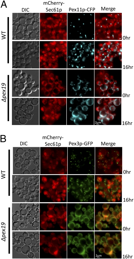
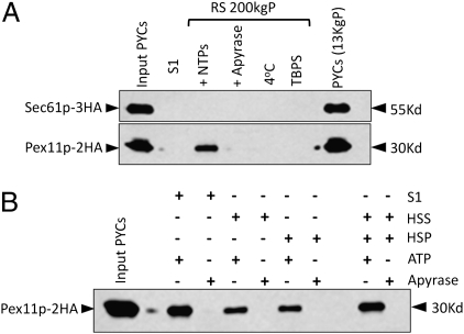
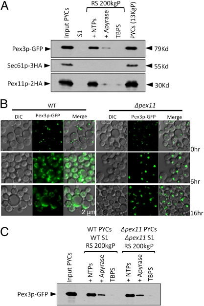
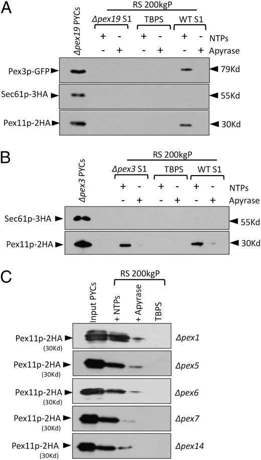
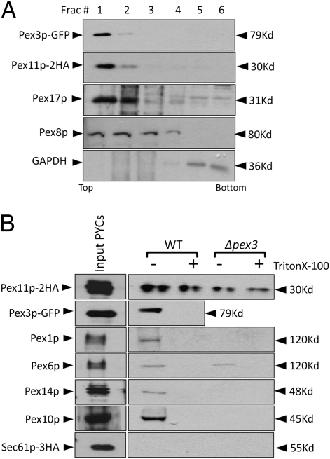
Several yeast and mammalian peroxisomal membrane proteins (PMPs) are delivered to peroxisomes via the endoplasmic reticulum (ER). Fluorescence microscopy showed a focused assembly of PMPs in a specialized domain of the ER, referred to as the preperoxisomal ER. It is proposed that preperoxisomal vesicles containing PMPs bud from this domain to either fuse with preexisting peroxisomes or to mature into functional peroxisomes by uptake of peroxisomal membrane and matrix proteins. However, such vesicular entities are not identified nor are the biochemical requirements for the budding process known. We developed an in vitro cell-free ER-budding assay using Pichia pastoris and followed two endogenous PMPs, Pex11p and Pex3p during their ER exit. Both the PMPs were copackaged in the ER-budded vesicles that float on a Nycodenz gradient. PMP budding from the ER was dependent on ATP, temperature, cytosol, and Pex19p and generated preperoxisomal vesicles with an incomplete complement of PMPs. Surprisingly, Pex11p budding was independent of Pex3p; however, the budded vesicles were devoid of most of the PMPs otherwise present in the wild-type vesicles and might represent peroxisomal remnants. Our findings provide a biochemical platform to uncover the mechanism of PMP budding from the ER.


Conflict of interest statement

The authors declare no conflict of interest.

Figures

Fig. 1.
Fig. 1.
Pex11p localization in WT and Δpex19 cells. Fluorescence microscopy analysis of oleate-grown WT and Δpex19 cells coexpressing the relevant proteins from PGAPPEX11CFP or PPEX3PEX3GFP and PSEC61mCherrySEC61. Cells were grown on YPD and switched during exponential phase to oleate medium for 0 or 16 h. mCherry–Sec61p (ER marker) localizes to punctate structures at the peripheral and nuclear ER. (A) In WT cells, Pex11p–CFP or Pex3p–GFP was partially localized with the Sec61p-labeled ER (0 h) and subsequently were found on the mature peroxisome cluster (16 h in oleate medium). (B) However, in Δpex19 cells Pex11p–CFP or Pex3p–GFP was mislocalized near the cell periphery partially associated with mCherry–Sec61p in the peripheral ER. No nonlinear adjustments or changes to gamma settings were made in the images.
Fig. 2.
Fig. 2.
Cell-free in vitro assay for Pex11p–2HA budding from the ER. (A) PYCs prepared from WT PPY12 cells expressing Pex11p–2HA and Sec61p–3HA were incubated with the WT S1 fraction for 90 min at 20 °C (lanes 3, 4, and 6) or 4 °C (lane 5) in the presence of an ATP-regenerating system (lanes 3, 5, and 6) or apyrase (lane 4). At the end of the budding reaction, samples were centrifuged at 13,000 rpm for 1 min to separate supernatants from the PYC pellet (lane 7), which still had the majority of the Pex11p–2HA and Sec61p–3HA. The reaction supernatant (RS) was centrifuged again at 200,000 × g and the pellet (RS 200 KgP) was resuspended in TBPS and analyzed here. PYCs in lane 1 represents nearly 3% load of the starting PYCs. (B) Soluble cytoplasmic (HSS) and the high-speed pelletable (HSP) membrane fractions obtained after further fractionating S1 were compared independently and together for their potential to support budding of Pex11p–2HA from the ER.
Fig. 3.
Fig. 3.
Division of preexisting peroxisomes is not the primary source of budded vesicles. (A) ER-budding assay was performed with WT cells coexpressing Pex11p–2HA, Sec61p–3HA, and Pex3p–GFP. Lane 1 represents nearly 3% load of the starting PYCs. (B) Fluorescence microscopy analysis of oleate-grown WT cells and Δpex11 cells expressing Pex3p–GFP. Cells were grown on YPD and switched during exponential phase to oleate medium. In WT cells, Pex3p–GFP labeled proliferating peroxisomes forming numerous punctate structures. The Δpex11 cells showed a severe block in peroxisome division even after growth for 16 h on oleate medium, resulting in bigger peroxisomes compared with those in WT cells. (C) ER-budding assay performed with WT cells and Δpex11 cells expressing Pex3p–GFP. Lane 1 represents nearly 3% load of the starting PYCs.
Fig. 4.
Fig. 4.
Pex19p is required for the budding of peroxisomal vesicles from the ER. (A) ER-budding assay was performed with Δpex19 cells coexpressing Pex11p–2HA, Sec61p–3HA, and Pex3p–GFP with either Δpex19 or WT cytosol. The expression of Pex11p–2HA was markedly low in Δpex19 and Δpex3 cells, so RS fractions of five budding reactions were pooled and analyzed together to obtain comparable levels of the protein. Neither Pex11p–2HA nor Pex3p–GFP was detected in the RS 200 KgP when Δpex19 cytosol was used. The budding of Pex11p–2HA was restored with WT cytosol (WT S1) in an ATP-dependent manner. Likewise, Pex3p–GFP budding too was restored with the WT cytosol (WT S1). PYCs in lane 1 represent nearly 3% load of the starting PYCs. (B) A similar ER-budding assay was performed with Δpex3 cells coexpressing Pex11p–2HA and Sec61p–3HA with Δpex3 or WT cytosol. Pex11p–2HA was detected in the RS 200-KgP fraction, indicating that Pex3p is not required for budding of preperoxisomal vesicles. Further addition of WT cytosol did not increase the budding of Pex11p–2HA vesicles. (C) The ER-budding assay was performed with Δpex1, Δpex5, Δpex6, Δpex7, and Δpex14 cells expressing Pex11p–2HA with their respective cytosols. Pex11p–2HA was detected in the RS 200 KgP in all of the mutants.
Fig. 5.
Fig. 5.
Pex3p and Pex11p budded from PYCs are membrane associated and are copackaged in the same vesicles. (A) The supernatant (RS) of the in vitro budding reaction was analyzed using a flotation gradient as described in Materials and Methods. Fractions (50 μL each) were collected from the top and analyzed by immunoblotting. Both the peroxins were found associated with buoyant membranes. (B) The RS fractions of 10 in vitro budding assays performed with WT and Δpex3 PYCs were pooled (900 μL) and spun at 200,000 × g. The pellet was resuspended in TBPS with or without Triton X-100 (1% vol/vol) and incubated with 80 μL of HA-affinity matrix to capture Pex11p–2HA-associated membranes. The incubation was for 6 h at 4 °C. The beads were washed three times, resuspended in 100 μL of TBPS, and analyzed.

References

    1. Fujiki Y, Rachubinski RA, Lazarow PB. Synthesis of a major integral membrane polypeptide of rat liver peroxisomes on free polysomes. Proc Natl Acad Sci USA. 1984;81:7127–7131. - PMC - PubMed
    1. Rachubinski RA, Fujiki Y, Mortensen RM, Lazarow PB. Acyl-Coa oxidase and hydratase-dehydrogenase, two enzymes of the peroxisomal β-oxidation system, are synthesized on free polysomes of clofibrate-treated rat liver. J Cell Biol. 1984;99:2241–2246. - PMC - PubMed
    1. Fagarasanu A, Mast FD, Knoblach B, Rachubinski RA. Molecular mechanisms of organelle inheritance: Lessons from peroxisomes in yeast. Nat Rev Mol Cell Biol. 2010;11:644–654. - PubMed
    1. Fagarasanu A, Fagarasanu M, Rachubinski RA. Maintaining peroxisome populations: A story of division and inheritance. Annu Rev Cell Dev Biol. 2007;23:321–344. - PubMed
    1. Motley AM, Hettema EH. Yeast peroxisomes multiply by growth and division. J Cell Biol. 2007;178:399–410. - PMC - PubMed

Publication types

MeSH terms

LinkOut - more resources