. 2011 May 31;108(22):9226-31.
doi: 10.1073/pnas.1100629108. Epub 2011 May 16.

Repair of injured proximal tubule does not involve specialized progenitors

Affiliations

Repair of injured proximal tubule does not involve specialized progenitors

Benjamin D Humphreys et al. Proc Natl Acad Sci U S A. .

Abstract

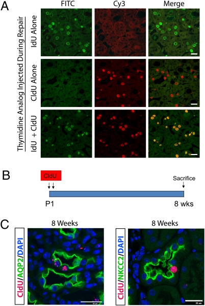
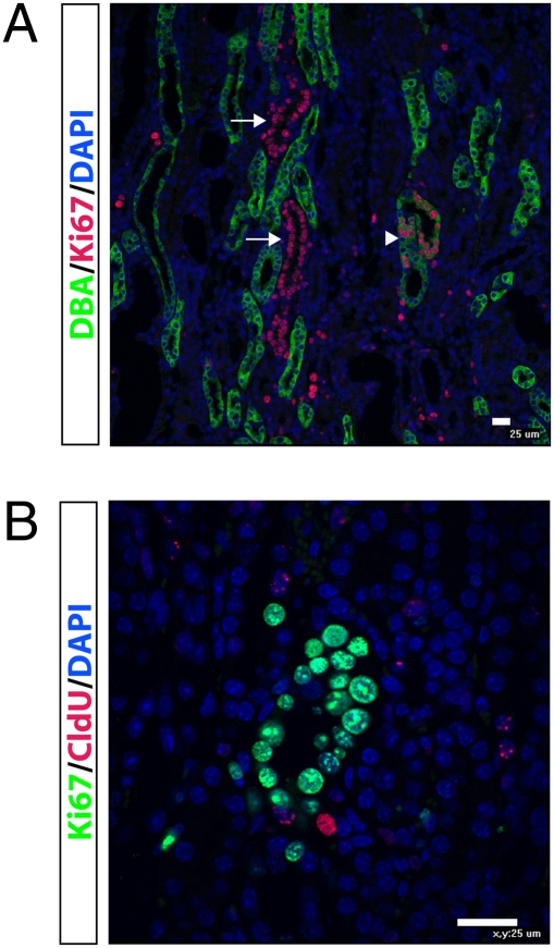
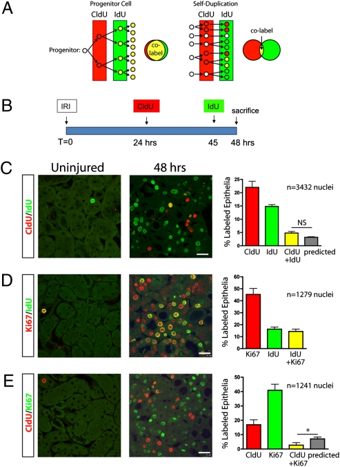
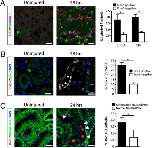
Recently we have established that the kidney tubular epithelium is repaired by surviving epithelial cells. It is not known, however, whether a population of intratubular adult progenitor cells are responsible for this epithelial repair after acute kidney injury. In this study, we used an unbiased DNA analog-based approach that does not rely on candidate markers to track multiple rounds of cell division in vivo. In the proximal tubule, robust thymidine analog incorporation was observed postinjury. Cell division was stochastic and enriched among cells that were injured and dedifferentiated. There was no evidence for the presence of a population of specialized progenitors that repeatedly divide in response to injury. Instead, these results indicate that after injury, new epithelial cells arise from self-duplication of surviving cells, most of which are injured. Because the renal papilla contains DNA label-retaining cells and has been proposed as a stem cell niche, we examined the proliferative behavior of these putative progenitors after ischemia-reperfusion injury. Although label-retaining cells in the renal papilla diminished with time after ischemia-reperfusion injury, they neither proliferated nor migrated to the outer medulla or cortex. Thus, nonlethally injured cells repopulate the kidney epithelium after injury in the absence of any specialized progenitor cell population.


Conflict of interest statement

The authors declare no conflict of interest.

Figures

Fig. 1.
Fig. 1.
Thymidine analog detection after either IRI or pulse chase. (A) To validate the specificity of thymidine analog labeling, IdU and CldU was injected alone or in combination 3 h before killing during repair. Kidney sections from outer medulla were probed with both anti-IdU (green) and anti-CldU (red) antibodies. When either IdU or CldU was injected alone there was no cross-reactivity, and when both IdU and CldU were injected together, there was concordant nuclear staining. (B) Newborn mice at P1 and P2 were injected with CldU followed by an 8-wk chase, to identify slow cycling cells in kidney. (Scale bar, 25 μm.) (C) LRCs were located in papilla, primarily in epithelial cells. ∼80% of LRC were AQP2-positive collecting duct cells, with the rest from other tubule segments such as NKCC2-positive thick ascending limb. (Scale bar, 25 μm.)
Fig. 2.
Fig. 2.
Papillary LRCs do not proliferate or migrate after IRI. (A) Groups of newborn mice (n = 3 per group) were injected with CldU at P1 and P2 and chased for 8 wk. Then, unilateral IRI was performed, and mice were killed at either 12, 24, 36, or 48 h after injury. IdU was injected 3 h before killing in each condition. (B) In the outer medulla, IdU (green) incorporation began 24 h after injury and increased thereafter. There were very rare CldU (red) positive cells in the outer medulla, and they neither incorporated IdU nor increased in number over time. In papilla, CldU-positive LRCs did not incorporate IdU during repair, although several IdU-positive cells could be found in papilla beginning 24 h after injury (arrows). (Scale bar, 25 μm.) (C) The percentage of LRCs in the cortex did not change during repair, indicating that there was no migration of LRCs from papilla to cortex. In contrast, the percentage of LRCs in the papilla decreased over the 48-h time course. *P = 0.003, 0 h compared with 48 h. (D) There was a dramatic increase in IdU-positive epithelial cells in the cortex, but only few cells in papilla divided after IRI. *P < 0.05, **P < 0.01, cortex compared with papilla. (E) Using the cell-cycle marker Ki67, there was a dramatic increase in cycling cells in cortex during repair, but very few Ki67-positive cells in renal papilla. *P < 0.05, **P < 0.01, cortex compared with papilla.
Fig. 3.
Fig. 3.
Chains of proliferating cells in the upper papilla do not derive from LRCs. (A) Forty-eight hours after IRI, tubules can be seen in which nearly all epithelial cell nuclei are Ki67-positive (red). These tubules are located in the base of the papilla, adjacent to the inner medulla. Some of these proliferating tubules are DBA-positive, indicating a collecting duct (arrowhead), and others are DBA-negative (arrows). (B) Costaining of CldU (red) and Ki67 (green) shows that LRCs are not contributing to the Ki67-positive cells, even though CldU-positive LRCs can be found adjacent to proliferating tubules. (Scale bar, 25 μm.)
Fig. 4.
Fig. 4.
Proximal tubule repair does not involve an intratubular progenitor population. (A) If an intratubular progenitor cell generates new epithelial cells during repair, such progenitors would selectively proliferate after injury resulting in a high percentage of colabeled nuclei after sequential CldU and IdU pulses. If all surviving epithelial cells are capable of proliferating through self-duplication, then cell division is stochastic and a certain fraction of cells will incorporate CldU, and a different fraction epithelial cells will incorporate IdU, with few cells incorporating both labels. (B) Adult mice were subject to unilateral IRI, and injected with CldU 24 h after injury and IdU 3 h before being killed at 48 h after injury. (C) Sham-operated kidneys showed little CldU (red) or IdU (green) uptake, but injured kidneys had substantial CldU and IdU uptake. A smaller fraction of cells was positive for both CldU and IdU, indicating sequential cell division in the prior 24 h, and this fraction was not different from that predicted if cell division were stochastic (%predicted = %CldU-positive × %IdU-positive). (D) Nearly all cells that incorporated IdU 3 h before being killed were also Ki67-positive, as expected. (E) The number of Ki67-positive cells was more than double the number of CldU-positive cells; however, very few cells were positive for both CldU and Ki67, and this number was significantly less than that predicted if cell division were stochastic. *P < 0.05. n = 3 mice for each condition. (Scale bar, 25 μm.)
Fig. 5.
Fig. 5.
Cell division occurs primarily in injured, dedifferentiated proximal tubule epithelial cells. (A) Mice treated with sequential CldU and IdU pulses and killed 48 h after injury show little thymidine analog uptake and no Kim-1 staining in sham-operated kidneys. In injured kidneys, there was strong apical Kim-1 staining. Tubules that were positive for Kim-1 were more likely to have divided over the previous 24 h (CldU) or 3 h (IdU), indicating that injured tubules are more likely to proliferate during repair. *P < 0.01. n = 3 mice for each condition. (B) Injury induces nuclear Pax2 expression in outer medullary tubular epithelia. Ki67-positive cycling epithelia were more likely to express the dedifferentiation marker Pax2. *P < 0.001. n = 4 mice for each condition. (C) Mice treated with a single pulse of BrdU before being killed 24 h after injury show reduced or absent basolateral Na/K/ATPase staining in the majority of BrdU-positive cells, indicating that proliferating cells are dedifferentiated at this time point. *P < 0.05. n = 3 mice for each condition. (Scale bars, 25 μm.)

References

    1. Messier B, Leblond CP. Cell proliferation and migration as revealed by radioautography after injection of thymidine-H3 into male rats and mice. Am J Anat. 1960;106:247–285. - PubMed
    1. Venkatachalam MA, Bernard DB, Donohoe JF, Levinsky NG. Ischemic damage and repair in the rat proximal tubule: Differences among the S1, S2, and S3 segments. Kidney Int. 1978;14(1):31–49. - PubMed
    1. Witzgall R, Brown D, Schwarz C, Bonventre JV. Localization of proliferating cell nuclear antigen, vimentin, c-Fos, and clusterin in the postischemic kidney. Evidence for a heterogenous genetic response among nephron segments, and a large pool of mitotically active and dedifferentiated cells. J Clin Invest. 1994;93:2175–2188. - PMC - PubMed
    1. Little MH, Bertram JF. Is there such a thing as a renal stem cell? J Am Soc Nephrol. 2009;20:2112–2117. - PubMed
    1. Elger M, et al. Nephrogenesis is induced by partial nephrectomy in the elasmobranch Leucoraja erinacea. J Am Soc Nephrol. 2003;14:1506–1518. - PubMed

Publication types

LinkOut - more resources