. 2011 May 31;108(22):9101-6.
doi: 10.1073/pnas.1016535108. Epub 2011 May 16.

Structural topology of phospholamban pentamer in lipid bilayers by a hybrid solution and solid-state NMR method

Affiliations

Structural topology of phospholamban pentamer in lipid bilayers by a hybrid solution and solid-state NMR method

Raffaello Verardi et al. Proc Natl Acad Sci U S A. .

Abstract

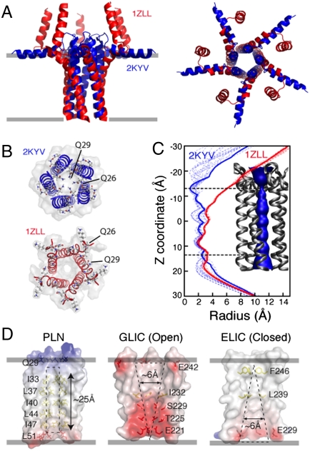
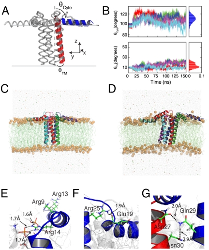
Phospholamban (PLN) is a type II membrane protein that inhibits the sarcoplasmic reticulum Ca(2+)-ATPase (SERCA), thereby regulating calcium homeostasis in cardiac muscle. In membranes, PLN forms pentamers that have been proposed to function either as a storage for active monomers or as ion channels. Here, we report the T-state structure of pentameric PLN solved by a hybrid solution and solid-state NMR method. In lipid bilayers, PLN adopts a pinwheel topology with a narrow hydrophobic pore, which excludes ion transport. In the T state, the cytoplasmic amphipathic helices (domains Ia) are absorbed into the lipid bilayer with the transmembrane domains arranged in a left-handed coiled-coil configuration, crossing the bilayer with a tilt angle of approximately 11° with respect to the membrane normal. The tilt angle difference between the monomer and pentamer is approximately 13°, showing that intramembrane helix-helix association forces dominate over the hydrophobic mismatch, driving the overall topology of the transmembrane assembly. Our data reveal that both topology and function of PLN are shaped by the interactions with lipids, which fine-tune the regulation of SERCA.


Conflict of interest statement

The authors declare no conflict of interest.

Figures

Fig. 1.
Fig. 1.
Multidimensional solution and solid-state NMR spectra defining the structural topology of PLN in DPC micelles, planar lipid bilayers, and lipid vesicles. (A) Two-dimensional planes from a 3D [1H, 1H,15N]-NOESY-HSQC spectrum (300-ms mixing time) showing NOEs between amide protons of PLN and methyl and methylene groups of DPC. Note the peaks at approximately 4.5 ppm are exchange cross-peaks between the protein amide resonances and the water signal. (B) Two-dimensional planes from 3D [1H, 1H,15N]-NOESY-HSQC (400-ms mixing time) on a mixed PLN sample with 1/1 of formula image 
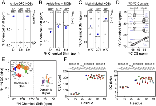
PLN. (C) Two-dimensional planes from 3D [1H, 13C, 13C]-HSQC-NOESY-HSQC experiment performed on a sample containing 1/1 of formula image and formula image PLN. (D) Two-dimensional-DARR experiment (200-ms mixing time) on a 1/1 sample of U-13C-Leu/U-13C-Ile PLN. Intraresidue and interprotomer cross-peaks are labeled in black and blue, respectively. (E) Overlay of 2D [1H, 15N]-PISEMA spectra of selectively labeled PLN in oriented DOPC/DOPE (4/1) lipid bilayers. (F) Plot of the CSA (Left) and DC (Right) values versus residue.
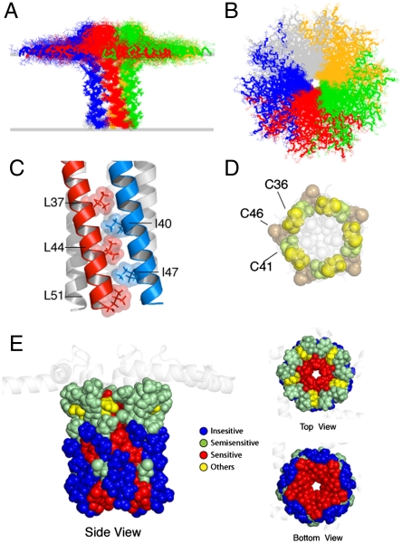
Fig. 2.
Fig. 2.
Pinwheel architecture of pentameric PLN in lipid bilayers. (A and B) PLN hybrid conformational ensemble. Overlay of the 20 lowest energy structures. The conformers were aligned using heavy atoms from residues 24 to 48. See Table S1 for structural statistics. (C) Domain II Leu/Ile zipper motif. Residues are shown using space filling model with atomic radii. (D) Spatial arrangement of the three cysteines in the PLN TM domain. The hydrophobic residues lining the inner pore are shown in light gray. Cys-41 and Cys-36 are at the interface between protomers. (E) Mapping of the mutagenesis studies on the PLN pentamer. Residues sensitive to mutation and oligomerization (1) are indicated in red. (Left) Side view of the PLN pentamer with residues from 24 to 52 shown in space filling model.
Fig. 3.
Fig. 3.
Comparison between 2KYV and 1ZLL structural models. (A) Overlay of the TM regions of the 1ZLL (red) and 2KYV (blue) models. Protein backbones were aligned using heavy atoms for residues 24 to 48. (B) Top view of the 1ZLL and 2KYV models. (C) Width of the central pore calculated with the program HOLE2 (68) for the 2KYV (blue dashed traces) and 1ZLL (red dashed traces) ensembles. Thick solid lines refer to the structures shown in A. (Inset) Surface rendering of the pore for the 2KYV model. (D) Electrostatic surface potentials of two pentameric ligand-gated ion channels (ELIC and GLIC) (44, 45) and PLN (2KYV).
Fig. 4.
Fig. 4.
MD simulations of PLN in explicit DOPC lipids. (A) Average structure of PLN from the hybrid NMR conformational ensemble with the TM domain (red) tilted by 11°. (B) Time course of the tilt angles of both the TM and cytoplasmic helices of PLN pentamer. (C and D) Snapshots of PLN from the MD trajectories taken at 10 and 150 ns. (E) Orientation of the three Arg residues in domain Ia. The guanidinium group of Arg-14 forms transient hydrogen bonds with several DOPC lipid molecules. (F) Arg-25 and Glu-19 salt bridge persistent throughout the MD trajectory. (G) Interprotomer contacts between the side chain of Gln-29, Asn-27, and Asn-30.

References

    1. MacLennan DH, Kranias EG. Phospholamban: A crucial regulator of cardiac contractility. Nat Rev Mol Cell Biol. 2003;4:566–577. - PubMed
    1. Simmerman HK, Jones LR. Phospholamban: Protein structure, mechanism of action, and role in cardiac function. Physiol Rev. 1998;78:921–47. - PubMed
    1. Zamoon J, Mascioni A, Thomas DD, Veglia G. NMR solution structure and topological orientation of monomeric phospholamban in dodecylphosphocholine micelles. Biophys J. 2003;85:2589–2598. - PMC - PubMed
    1. Traaseth NJ, et al. Structural and dynamic basis of phospholamban and sarcolipin inhibition of ca(2+)-ATPase. Biochemistry. 2008;47:3–13. - PMC - PubMed
    1. Haghighi K, et al. A mutation in the human phospholamban gene, deleting arginine 14, results in lethal, hereditary cardiomyopathy. Proc Natl Acad Sci USA. 2006;103:1388–1393. - PMC - PubMed

Publication types

LinkOut - more resources