

Loss of nuclear localized and tyrosine phosphorylated Stat5 in breast cancer predicts poor clinical outcome and increased risk of antiestrogen therapy failure
- PMID: 21576635
- PMCID: PMC3675698
- DOI: 10.1200/JCO.2010.30.3552
Loss of nuclear localized and tyrosine phosphorylated Stat5 in breast cancer predicts poor clinical outcome and increased risk of antiestrogen therapy failure
Abstract
Purpose: To investigate nuclear localized and tyrosine phosphorylated Stat5 (Nuc-pYStat5) as a marker of prognosis in node-negative breast cancer and as a predictor of response to antiestrogen therapy.
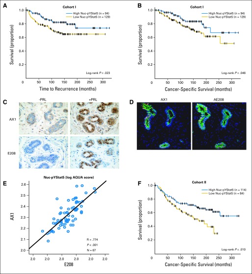
Patients and methods: Levels of Nuc-pYStat5 were analyzed in five archival cohorts of breast cancer by traditional diaminobenzidine-chromogen immunostaining and pathologist scoring of whole tissue sections or by immunofluorescence and automated quantitative analysis (AQUA) of tissue microarrays.
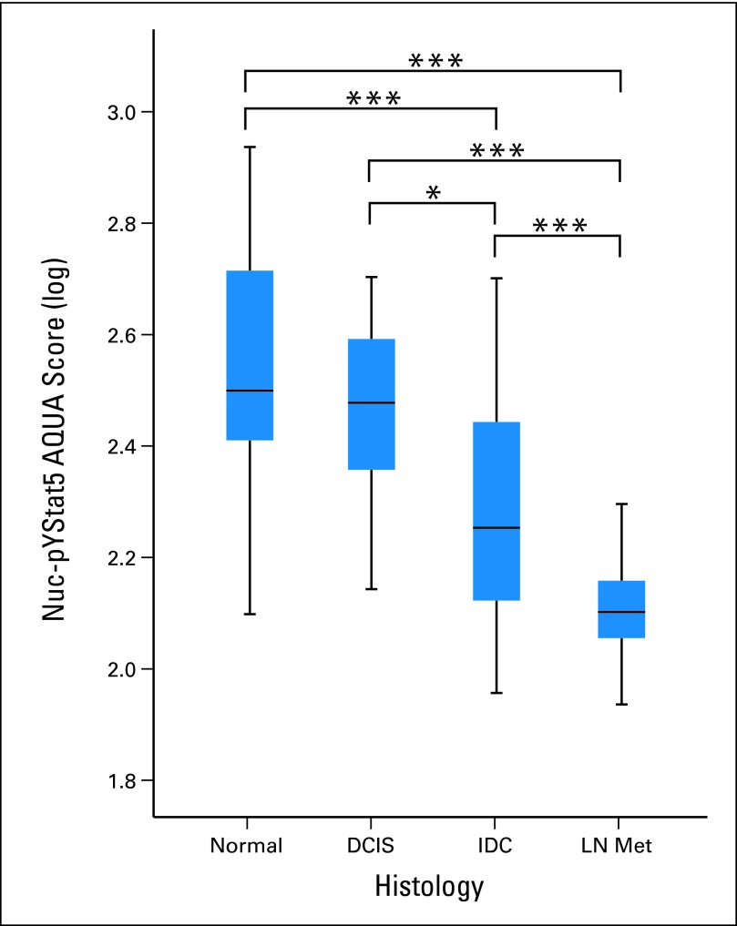
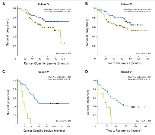
Results: Nuc-pYStat5 was an independent prognostic marker as measured by cancer-specific survival (CSS) in patients with node-negative breast cancer who did not receive systemic adjuvant therapy, when adjusted for common pathology parameters in multivariate analyses both by standard chromogen detection with pathologist scoring of whole tissue sections (cohort I; n = 233) and quantitative immunofluorescence of a tissue microarray (cohort II; n = 291). Two distinct monoclonal antibodies gave concordant results. A progression array (cohort III; n = 180) revealed frequent loss of Nuc-pYStat5 in invasive carcinoma compared to normal breast epithelia or ductal carcinoma in situ, and general loss of Nuc-pYStat5 in lymph node metastases. In cohort IV (n = 221), loss of Nuc-pYStat5 was associated with increased risk of antiestrogen therapy failure as measured by univariate CSS and time to recurrence (TTR). More sensitive AQUA quantification of Nuc-pYStat5 in antiestrogen-treated patients (cohort V; n = 97) identified by multivariate analysis patients with low Nuc-pYStat5 at elevated risk for therapy failure (CSS hazard ratio [HR], 21.55; 95% CI, 5.61 to 82.77; P < .001; TTR HR, 7.30; 95% CI, 2.34 to 22.78; P = .001). CONCLUSION Nuc-pYStat5 is an independent prognostic marker in node-negative breast cancer. If confirmed in prospective studies, Nuc-pYStat5 may become a useful predictive marker of response to adjuvant hormone therapy.
Conflict of interest statement
Authors' disclosures of potential conflicts of interest and author contributions are found at the end of this article.
Figures
Comment in
-
Stat5: from breast development to cancer prognosis, prediction, and progression.J Clin Oncol. 2011 Jun 20;29(18):2443-4. doi: 10.1200/JCO.2010.34.2014. Epub 2011 May 16. J Clin Oncol. 2011. PMID: 21576641 No abstract available.
-
Prognostic or predictive? It's time to get back to definitions!J Clin Oncol. 2011 Dec 10;29(35):4718; author reply 4718-9. doi: 10.1200/JCO.2011.38.3729. Epub 2011 Oct 31. J Clin Oncol. 2011. PMID: 22042948 No abstract available.
References
-
- Liu X, Robinson GW, Wagner KU, et al. Stat5a is mandatory for adult mammary gland development and lactogenesis. Genes Dev . 1997;11:179–186. - PubMed
-
- Teglund S, McKay C, Schuetz E, et al. Stat5a and Stat5b proteins have essential and nonessential, or redundant, roles in cytokine responses. Cell . 1998;93:841–850. - PubMed
-
- Xie J, LeBaron MJ, Nevalainen MT, et al. Role of tyrosine kinase Jak2 in prolactin-induced differentiation and growth of mammary epithelial cells. J Biol Chem . 2002;277:14020–14030. - PubMed
Publication types
MeSH terms
Substances
Grants and funding
LinkOut - more resources
Full Text Sources
Medical
Research Materials
Miscellaneous
