Case Reports
. 2011 May 17;76(20):1705-10.
doi: 10.1212/WNL.0b013e31821a44f1.

A case of multiple sclerosis presenting with inflammatory cortical demyelination

Affiliations
Case Reports

A case of multiple sclerosis presenting with inflammatory cortical demyelination

B F Gh Popescu et al. Neurology. .

Abstract

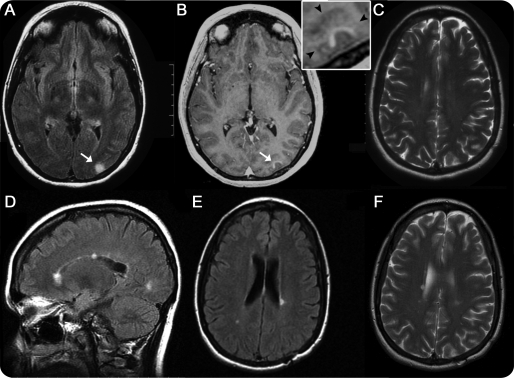
Objective: To describe a patient presenting with a clinically silent, incidentally found, and pathologically confirmed active demyelinating solitary cortical lesion showing MRI gadolinium contrast enhancement, in whom biopsy was performed before the radiographic appearance of disseminated white matter lesions.

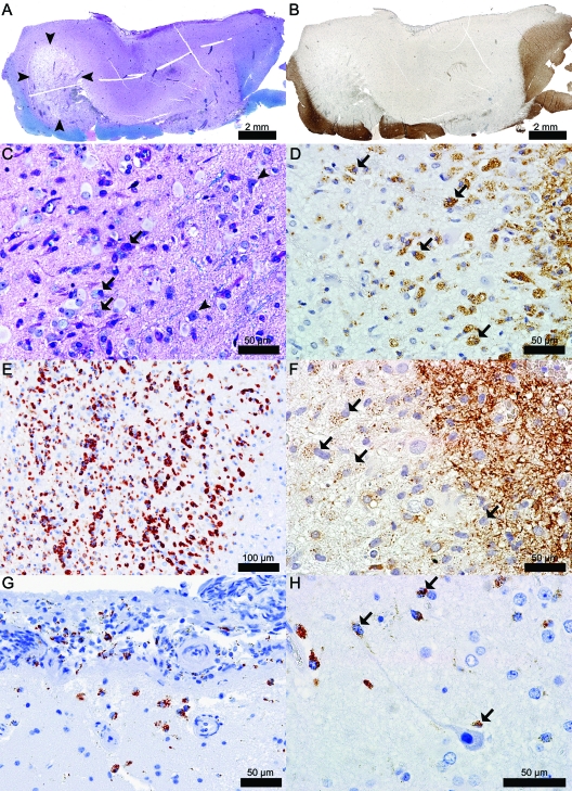
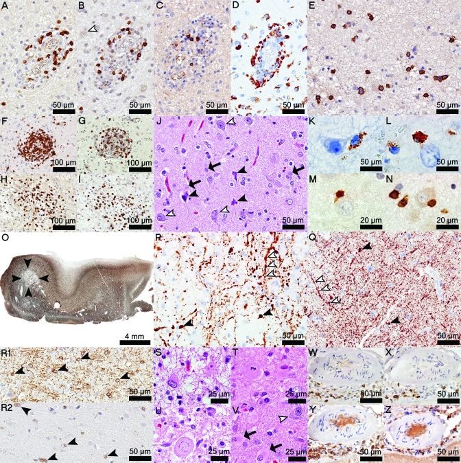
Methods: Neurologic examination, MRI, CSF and serologic analyses, and brain biopsy were performed. Sections of formalin-fixed paraffin-embedded biopsied brain tissue were stained with histologic and immunohistochemical stains.

Results: Biopsy revealed an inflammatory subpial lesion containing lymphocytes and myelin-laden macrophages. Recurrent relapses with dissemination of MRI-typical white matter lesions characterized the subsequent course.

Conclusions: Our findings highlight that cortical demyelination occurs on a background of inflammation and suggest that the noninflammatory character of chronic cortical demyelination may relate to long intervals between lesion formation and autopsy. This case provides pathologic evidence of relapsing-remitting MS presenting with inflammatory cortical demyelination and emphasizes the importance of considering demyelinating disease in the differential diagnosis of patients presenting with a solitary cortical enhancing lesion.


Figures

Figure 1
Figure 1. Brain MRI prior to biopsy (A–C) and 69 months after biopsy (D–F)
(A) Axial fluid-attenuated inversion recovery (FLAIR) image of left occipital cortical lesion (arrow). (B) Axial T1-weighted image with contrast showing enhancement of the cortical lesion (arrow); inset shows that contrast enhancement is within the cortical gray matter (arrowheads indicate the gray/white matter junction). (C) Axial T2-weighted image showing no periventricular lesions. (D) Sagittal FLAIR image showing the appearance of multiple new white matter lesions, with 2 of the lesions involving the corpus callosum. (E) Axial FLAIR image with the appearance of new periventricular lesions. (F) Axial T2-weighted image at nearly corresponding level of image C showing the appearance of new periventricular white matter lesions.
Figure 2
Figure 2. Biopsy of the initial MRI enhancing lesion shown in figure 1 revealed a highly inflammatory subpial lesion with predominance of macrophages involved in active demyelination
(A) Subpial lesion showing loss of myelin and an area (arrowheads) of cavitary changes (Luxol fast blue [LFB]/periodic acid-Schiff [PAS]). (B) Subpial lesion showing loss of immunoreactivity for myelin specific proteins (proteolipid protein [PLP]). (C) Actively demyelinating macrophages (arrows); arrowheads indicate neurons (LFB/PAS). (D) Actively demyelinating macrophages (arrows) (PLP). (E) Foamy macrophages (KiM1P). (F) Actively demyelinating macrophages (arrows) (2′3′-cyclic-nucleotide 3′-phosphodiesterase [CNPase]). (G) Subpial and CSF foamy macrophages (KiM1P). (H) Macrophages/microglia (arrows) in close apposition to neurons and neurites (KiM1P).
Figure 3
Figure 3. Cortical and meningeal lymphocytic infiltration, axonal and neuronal injury, loss of oligodendrocytes, and reactive astrocytosis are other neuropathologic findings in this biopsy
(A) Perivascular CD3+ T cells (CD3). (B) Perivascular CD8+ T cells (CD8, white arrowhead shows a normal-appearing neuron). (C) Perivascular B cells (CD20). (D) Perivascular macrophages (KiM1P). (E) Parenchymal plasma cells (CD138). (F) Junctional perivascular CD3+ T cells (CD3). (G) Junctional perivascular CD8+ T cells (CD8). (H) Junctional parenchymal CD3+ T cells (CD3). (I) Junctional parenchymal CD8+ T cells (CD8). (J) Pyknotic neurons (black arrowheads) and reactive astrocytes (arrows) scattered among healthy neurons (white arrowheads) (hematoxylin & eosin [H&E]). (K, L) Macrophages in close apposition to neurons (KiM1P). (M) CD3+ T cells (CD3) and (N) CD8+ T cells (CD8) in close apposition to neurons. (O) Good axonal preservation, except in the cavitated area (arrowheads) (neurofilament [NF]). (P) Axonal swellings in the cavitated area (black arrowheads); segmental axonal swellings are seen along neurites (white arrowheads) (NF). (Q) Axonal swellings in normal-appearing gray matter (black arrowheads); white arrowheads follow a neurite with segmental axonal swellings (NF). (R1) Oligodendrocytes in normal-appearing cortex (2′3′-cyclic-nucleotide 3′-phosphodiesterase [CNPase]). (R2) Loss of oligodendrocytes in the subpial lesion (CNPase). (S) Gemistocytes (H&E). (T) Mitotic astrocytes (H&E). (U) Creutzfeldt cells (H&E). (V) Cortical reactive astrocytosis (arrows) extending beyond the lesion borders; healthy neuron (white arrowhead) (H&E). (W) Meningeal CD3+ T cells (CD3). (X) Meningeal CD8+ T cells (CD8). (Y) Meningeal B cells (CD20). (Z) Meningeal plasma cells (CD138).

References

    1. Brownell B, Hughes JT. The distribution of plaques in the cerebrum in multiple sclerosis. J Neurol Neurosurg Psychiatry 1962;25:315–320 - PMC - PubMed
    1. Kidd D, Barkhof F, McConnell R, Algra PR, Allen IV, Revesz T. Cortical lesions in multiple sclerosis. Brain 1999;122:17–26 - PubMed
    1. Peterson JW, Bo L, Mork S, Chang A, Trapp BD. Transected neurites, apoptotic neurons, and reduced inflammation in cortical multiple sclerosis lesions. Ann Neurol 2001;50:389–400 - PubMed
    1. Kutzelnigg A, Lucchinetti CF, Stadelmann C, et al. Cortical demyelination and diffuse white matter injury in multiple sclerosis. Brain 2005;128:2705–2712 - PubMed
    1. Merkler D, Ernsting T, Kerschensteiner M, Bruck W, Stadelmann C. A new focal EAE model of cortical demyelination: multiple sclerosis-like lesions with rapid resolution of inflammation and extensive remyelination. Brain 2006;129:1972–1983 - PubMed

Publication types

MeSH terms