. 2010;7(4):173-88.
doi: 10.3233/CBM-2010-0188.

Molecular and functional imaging of invasion and metastasis: windows into the metastatic cascade

Affiliations

Molecular and functional imaging of invasion and metastasis: windows into the metastatic cascade

Ioannis Stasinopoulos et al. Cancer Biomark. 2010.

Abstract

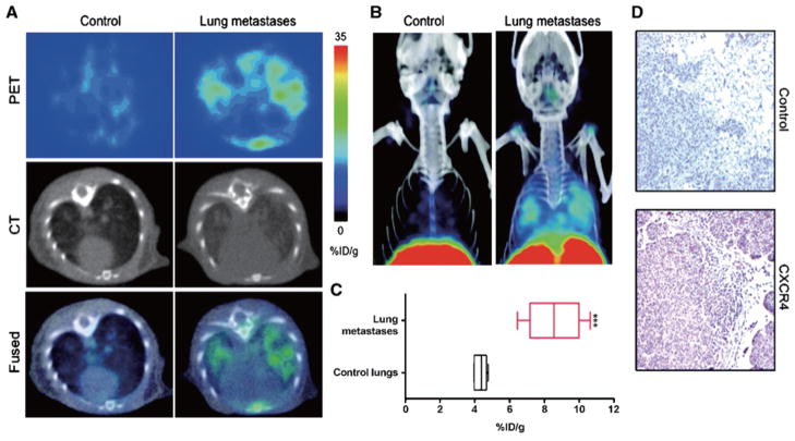
The ability of cancer cells to invade, metastasize, and form distant colonies, is one of the key characteristics that confers lethality to cancer. Metastatic cancer cells typically become refractory to treatment. The metastatic cascade is a multi-step process that is governed by events within the cancer cell, the tumor microenvironment, and the distant environments that are invaded and colonized by the cancer cells. Noninvasive imaging techniques are facilitating a close examination of the stepwise journey of the cancer cell from the primary tumor to the distant metastatic site. Here we have discussed the metastatic process, and how molecular and functional imaging of cancer are providing new insights into the metastatic cascade that can be exploited for treatment of metastatic disease.


Figures

Fig. 1
Fig. 1
Stages of Metastatic Progression. Metastasis proceeds through the progressive acquisition of traits that allow malignant cells originating in one organ to disseminate and colonize a secondary site. Although these traits are depicted as part of a contiguous biological sequence, their acquisition during metastatic progression need not follow this particular order. Although in some cases several factors may be necessary to implement a single step in this cascade, other mediators of metastasis may facilitate execution of multiple stages simultaneously. Similarly, the specific steps of this sequence that are rate limiting for metastatic progression may also vary from one tumor to the next. Reprinted with permission from [3].
Fig. 2
Fig. 2
Cancer cells in primary tumors are surrounded by a complex microenvironment comprising numerous cells including endothelial cells of the blood and lymphatic circulation, stromal fibroblasts and a variety of bone marrow-derived cells (BMDCs) including macrophages, myeloid-derived suppressor cells (MDSCs), TIE2-expressing monocytes (TEMs) and mesenchymal stem cells (MSCs). Reprinted with permission from [11].
Fig. 3
Fig. 3
(A) Schematic display of the sample structure (center) of a cell-perfusion assay, which can be identified clearly in the representative T1-weighted 1H MR image on the right. (B) T1-weighted 1H MR images and Invasion Indices over time demonstrating the effect of indomethacin treatment on degradation and invasion of ECM gel by MDA-MB-435 breast cancer cells. Values of the Invasion Indices are presented as mean ± SE. Reprinted with permission [56].
Fig. 4
Fig. 4
Schematic overview of the four essential steps of brain metastasis formation. 1. Arrest at blood vessels by size restriction. 2. Active extravasation though the holes of the vascular wall. 3. Localization at a perivascular position. 4. Macrometastasis either through continuous growth of cell clusters and vascular cooption, or by the formation of new tumors from single cells. Reprinted with permission from [69].
Fig. 5
Fig. 5
Additional value of CT in 18F-FHBG PET images of metastasis. (A) Thirty-five days after intraventricular injection of 1.5 × 10⁶ A375M-3F melanoma cells in nude mouse, 18F-FHBG PET/CT allows precise anatomic localization of metastasis in interscapular fat (a), right eye (b), right humeral head (c), and left mandibula (d) as shown by green arrows. Lack of anatomic landmarks on PET alone is illustrated by white arrows. (B) BLI shows same lesions as seen on 18F-FHBG PET/CT but does not provide information on depth of lesion. (C) Ex vivo thymidine kinase and luciferase assays of lesions (1) and contralateral controls (2) validate imaging observations. Reprinted with permission from [92].
Fig. 6
Fig. 6
CXCR4 imaging of MDA-MB-231–derived lung metastases with [64Cu]AMD3100. NOD/SCID mice that received either 2 × 10⁶ MDA-MB-231 cells or HBSS received 11 MBq (300 μCi) of [64Cu]AMD3100 at 35 d after inoculation. Whole body images were acquired at 90 min after injection. A. Transaxial PET, CT, and fused sections of lung metastasis and control mice. B. Volume-rendered whole body image showing clear accumulation of radioactivity in the lung metastases. Top slices of the volume-rendered images were cut for clear visualization of lung uptake. C. Box-and-whisker plot of the biodistribution analysis of lungs from mice injected with 740 kBq of [64Cu]AMD3100 at 90 min post-injection. All radioactivity values were converted into %ID/g of tissue and are means ± SEM of four to five animals. D. Representative microscopy images of 10-μm-thick CXCR4-stained and control antibody–stained sections of the lung metastases obtained at × 10 magnification. Significance is indicated by asterisks (*), and the comparative reference are lungs from mice injected with Hanks balanced salt solution. ***P < 0.001. Reprinted with permission from [95].
Fig. 7
Fig. 7
A. Axial T2-weighted fast spin-echo image (TR (repetition time)/TE (echo time): 3100/135 milliseconds) at 3 T with combined body and spine matrix coils for localization of the enlarged lymph node (arrows). I, iliac bone; S, sigmoid; V, iliac vessels. B. The phase-corrected 1H-MR spectrum (without baseline correction) obtained from the TE-averaged and combined water and lipid-suppressed single voxel MR spectroscopy measurement. A peak at 4.7 ppm for residual water spins and a peak at about 3.2 ppm assigned to the methyl groups of choline-containing compounds (tCho) were observed. Note the absence of lipid signals from 1 to 2.5 ppm. The coronal reference image from the T1-weighted 3-dimensional gradient-echo sequence (TR/TE: 6.47/2.54 milliseconds) and the axial T2-weighted fast spin-echo image are shown to indicate the voxel placement. C. An axial spectral map of all voxels inside the volume-of-interest from the 1H-MR spectroscopic imaging measurement (TR/TE: 1500/100 milliseconds) in which the tCho resonances are underlined. Outside the lymph node, large lipid signals are present; the tCho signal is only present inside. D. Interpolated coronal and axial tCho color-coded maps constructed using the integral of a Gaussian fit to the tCho signal. Reprinted with permission from [101].

Similar articles

Cited by

References

    1. Dorudi S, Hart IR. Mechanisms underlying invasion and metastasis. Curr Opin Oncol. 1993;5:130–135. - PubMed
    1. Meyer T, Hart IR. Mechanisms of tumour metastasis. Eur J Cancer. 1998;34:214–221. - PubMed
    1. Gupta GP, Massague J. Cancer metastasis: building a framework. Cell. 2006;127:679–695. - PubMed
    1. Coghlin C, Murray GI. Current and emerging concepts in tumour metastasis. J Pathol. 2010;222:1–15. - PubMed
    1. Klein CA. Parallel progression of primary tumours and metastases. Nat Rev Cancer. 2009;9:302–312. - PubMed

Publication types

LinkOut - more resources