

Observation of nonspherical particle behaviors for continuous shape-based separation using hydrodynamic filtration
- PMID: 21584211
- PMCID: PMC3094456
- DOI: 10.1063/1.3580757
Observation of nonspherical particle behaviors for continuous shape-based separation using hydrodynamic filtration
Abstract
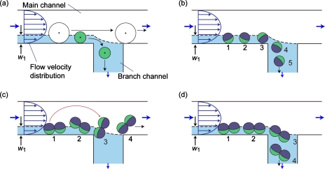
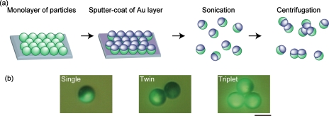
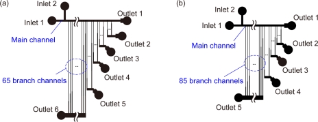
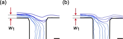
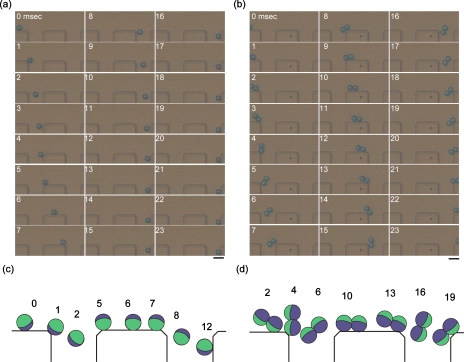
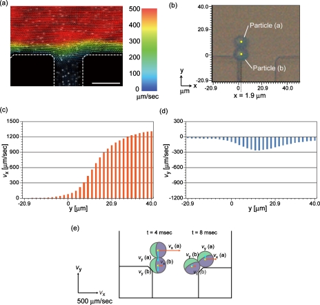
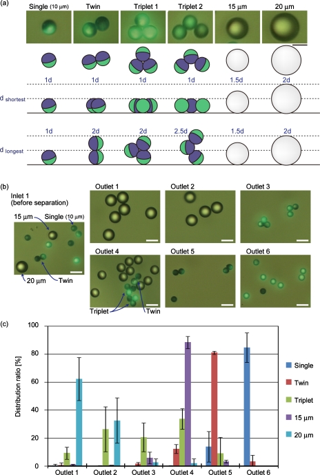
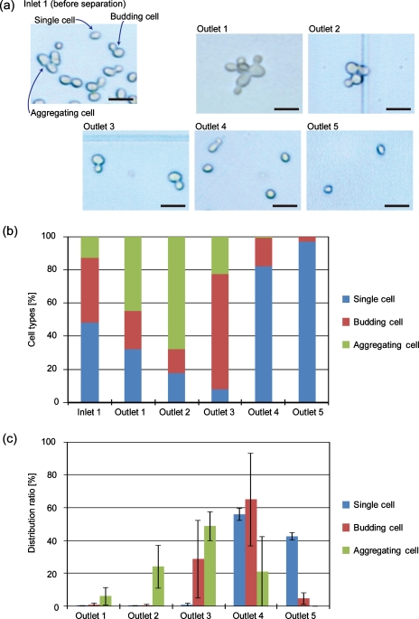
Selection of particles or cells of specific shapes from a complex mixture is an essential procedure for various biological and industrial applications, including synchronization of the cell cycle, classification of environmental bacteria, and elimination of aggregates from synthesized particles. Here, we investigate the separation behaviors of nonspherical and spherical particles∕cells in the hydrodynamic filtration (HDF) scheme, which was previously developed for continuous size-dependent particle∕cell separation. Nonspherical particle models were prepared by coating the hemisphere of spherical polymer particles with a thin Au layer and by bonding the Janus particles to form twins and triplets resembling dividing and aggregating cells, respectively. High-speed imaging revealed a difference in the separation behaviors of spherical and nonspherical particles at a branch point; nonspherical particles showed rotation behavior and did not enter the branch channel even when their minor axis was smaller than the virtual width of the flow region entering the branch channel, w(1). The confocal-laser high-speed particle intensity velocimetry system visualized the flow profile inside the HDF microchannel, demonstrating that the steep flow-velocity distribution at the branch point is the main factor causing the rotation behavior of nonspherical particles. As applications, we successfully separated spherical and nonspherical particles with various major∕minor lengths and also demonstrated the selection of budding∕single cells from a yeast cell mixture. We therefore conclude that the HDF scheme can be used for continuous shape-based particle∕cell separation.
Figures
Similar articles
-
Formation of Nonspherical Cellulose Acetate Microparticles under Microflow.Langmuir. 2024 Dec 31;40(52):27314-27322. doi: 10.1021/acs.langmuir.4c03430. Epub 2024 Dec 18. Langmuir. 2024. PMID: 39696796 Free PMC article.
-
Continuous particle separation in a microchannel having asymmetrically arranged multiple branches.Lab Chip. 2005 Jul;5(7):778-84. doi: 10.1039/b501885d. Epub 2005 May 19. Lab Chip. 2005. PMID: 15970972
-
Hydrodynamic filtration for on-chip particle concentration and classification utilizing microfluidics.Lab Chip. 2005 Nov;5(11):1233-9. doi: 10.1039/b509386d. Epub 2005 Sep 26. Lab Chip. 2005. PMID: 16234946
-
Current status and future developments in preparation and application of nonspherical polymer particles.Adv Colloid Interface Sci. 2018 Jun;256:126-151. doi: 10.1016/j.cis.2018.04.010. Epub 2018 Apr 20. Adv Colloid Interface Sci. 2018. PMID: 29705026 Review.
-
Transport of Non-Spherical Particles in Square Microchannel Flows: A Review.Micromachines (Basel). 2021 Mar 7;12(3):277. doi: 10.3390/mi12030277. Micromachines (Basel). 2021. PMID: 33800014 Free PMC article. Review.
Cited by
-
Enhanced Blood Plasma Extraction Utilising Viscoelastic Effects in a Serpentine Microchannel.Biosensors (Basel). 2022 Feb 14;12(2):120. doi: 10.3390/bios12020120. Biosensors (Basel). 2022. PMID: 35200380 Free PMC article.
-
Microfluidic electrical sorting of particles based on shape in a spiral microchannel.Biomicrofluidics. 2014 Jan 14;8(1):014101. doi: 10.1063/1.4862355. eCollection 2014 Jan. Biomicrofluidics. 2014. PMID: 24753722 Free PMC article.
-
High-throughput and clogging-free microfluidic filtration platform for on-chip cell separation from undiluted whole blood.Biomicrofluidics. 2016 Feb 12;10(1):014118. doi: 10.1063/1.4941985. eCollection 2016 Jan. Biomicrofluidics. 2016. PMID: 26909124 Free PMC article.
-
Hydrodynamic particle focusing design using fluid-particle interaction.Biomicrofluidics. 2013 Sep 11;7(5):54104. doi: 10.1063/1.4821170. eCollection 2013. Biomicrofluidics. 2013. PMID: 24404067 Free PMC article.
-
Shape-based separation of microalga Euglena gracilis using inertial microfluidics.Sci Rep. 2017 Sep 7;7(1):10802. doi: 10.1038/s41598-017-10452-5. Sci Rep. 2017. PMID: 28883551 Free PMC article.
References
-
- Bauman R. W., in Microbiology: With Diseases by Body System, 2nd ed., edited by Berriman L. (Pearson Benjamin Cummings, San Francisco, 2009), Chaps. 4 and 11.
-
- Fants P. and Brooks R., The Cell Cycle: A Practical Approach (IRL, Oxford, 1993), Chaps. 1, 2, and 4.
-
- Needham D., Cell Biophys. CBIODE 18, 99 (1991). - PubMed
LinkOut - more resources
Full Text Sources
Other Literature Sources
Molecular Biology Databases
Research Materials