

Three-dimensional echocardiography for left ventricular quantification: fundamental validation and clinical applications
- PMID: 21584798
- PMCID: PMC3189315
- DOI: 10.1007/s12471-011-0160-y
Three-dimensional echocardiography for left ventricular quantification: fundamental validation and clinical applications
Abstract
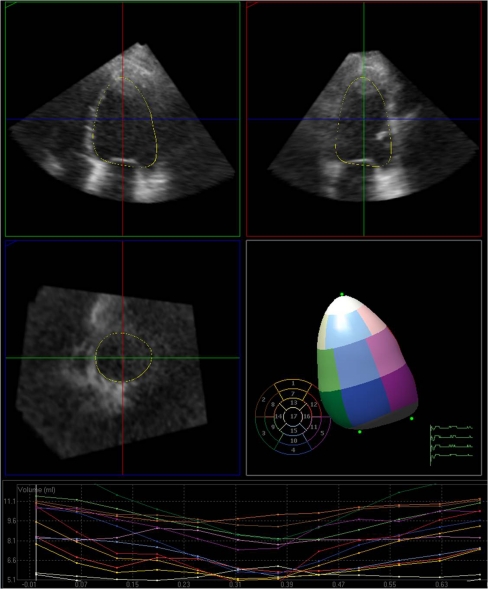
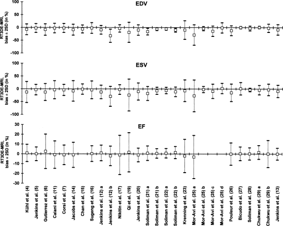
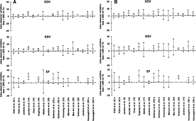
One of the earliest applications of clinical echocardiography is evaluation of left ventricular (LV) function and size. Accurate, reproducible and quantitative evaluation of LV function and size is vital for diagnosis, treatment and prediction of prognosis of heart disease. Early three-dimensional (3D) echocardiographic techniques showed better reproducibility than two-dimensional (2D) echocardiography and narrower limits of agreement for assessment of LV function and size in comparison to reference methods, mostly cardiac magnetic resonance (CMR) imaging, but acquisition methods were cumbersome and a lack of user-friendly analysis software initially precluded widespread use. Through the advent of matrix transducers enabling real-time three-dimensional echocardiography (3DE) and improvements in analysis software featuring semi-automated volumetric analysis, 3D echocardiography evolved into a simple and fast imaging modality for everyday clinical use. 3DE provides the possibility to evaluate the entire LV in three spatial dimensions during the complete cardiac cycle, offering a more accurate and complete quantitative evaluation the LV. Improved efficiency in acquisition and analysis may provide clinicians with important diagnostic information within minutes. The current article reviews the methodology and application of 3DE for quantitative evaluation of the LV, provides the scientific evidence for its current clinical use, and discusses its current limitations and potential future directions.
Figures
References
-
- Buck T, Hunold P, Wentz KU, et al. Tomographic three-dimensional echocardiographic determination of chamber size and systolic function in patients with left ventricular aneurysm: comparison to magnetic resonance imaging, cineventriculography, and two-dimensional echocardiography. Circulation. 1997;96:4286–4297. - PubMed
-
- Chuang ML, Hibberd MG, Salton CJ, et al. Importance of imaging method over imaging modality in noninvasive determination of left ventricular volumes and ejection fraction: assessment by two- and three-dimensional echocardiography and magnetic resonance imaging. J Am Coll Cardiol. 2000;35:477–484. doi: 10.1016/S0735-1097(99)00551-3. - DOI - PubMed
-
- Mannaerts HF, Van Der Heide JA, Kamp O, et al. Quantification of left ventricular volumes and ejection fraction using freehand transthoracic three-dimensional echocardiography: comparison with magnetic resonance imaging. J Am Soc Echocardiogr. 2003;16:101–109. doi: 10.1067/mje.2003.7. - DOI - PubMed
-
- Kuhl HP, Schreckenberg M, Rulands D, et al. High-resolution transthoracic real-time three-dimensional echocardiography: quantitation of cardiac volumes and function using semi-automatic border detection and comparison with cardiac magnetic resonance imaging. J Am Coll Cardiol. 2004;43:2083–2090. doi: 10.1016/j.jacc.2004.01.037. - DOI - PubMed
LinkOut - more resources
Full Text Sources
