

Hypoxia in the regulation of neural stem cells
- PMID: 21584807
- PMCID: PMC11115125
- DOI: 10.1007/s00018-011-0723-5
Hypoxia in the regulation of neural stem cells
Abstract
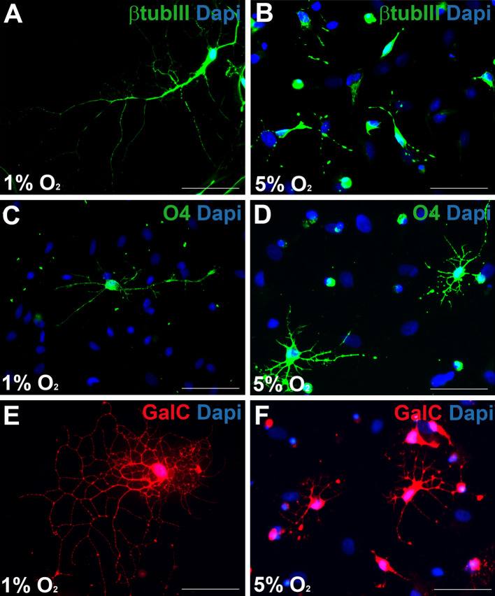
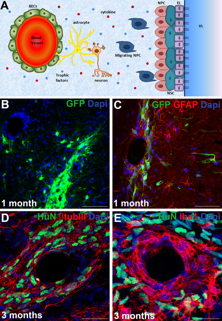
In aerobic organisms, oxygen is a critical factor in tissue and organ morphogenesis from embryonic development throughout post-natal life, as it regulates various intracellular pathways involved in cellular metabolism, proliferation, survival and fate. In the mammalian central nervous system, oxygen plays a critical role in regulating the growth and differentiation state of neural stem cells (NSCs), multipotent neuronal precursor cells that reside in a particular microenvironment called the neural stem cell niche and that, under certain physiological and pathological conditions, differentiate into fully functional mature neurons, even in adults. In both experimental and clinical settings, oxygen is one of the main factors influencing NSCs. In particular, the physiological condition of mild hypoxia (2.5-5.0% O(2)) typical of neural tissues promotes NSC self-renewal; it also favors the success of engraftment when in vitro-expanded NSCs are transplanted into brain of experimental animals. In this review, we analyze how O(2) and specifically hypoxia impact on NSC self-renewal, differentiation, maturation, and homing in various in vitro and in vivo settings, including cerebral ischemia, so as to define the O(2) conditions for successful cell replacement therapy in the treatment of brain injury and neurodegenerative diseases.
Figures

Similar articles
-
New Insights into the Role of Mild Hypoxia in Regulating Neural Stem Cell Characteristics.Stem Cells Dev. 2024 Jul;33(13-14):333-342. doi: 10.1089/scd.2024.0020. Epub 2024 Jun 11. Stem Cells Dev. 2024. PMID: 38753713 Review.
-
Time-sensitive effects of hypoxia on differentiation of neural stem cells derived from mouse embryonic stem cells in vitro.Neurol Res. 2014 Sep;36(9):804-13. doi: 10.1179/1743132814Y.0000000338. Epub 2014 Feb 11. Neurol Res. 2014. PMID: 24620986
-
In vitro investigation of the mechanism underlying the effect of ginsenoside on the proliferation and differentiation of neural stem cells subjected to oxygen-glucose deprivation/reperfusion.Int J Mol Med. 2018 Jan;41(1):353-363. doi: 10.3892/ijmm.2017.3253. Epub 2017 Nov 13. Int J Mol Med. 2018. PMID: 29138802 Free PMC article.
-
A Tale of Two: When Neural Stem Cells Encounter Hypoxia.Cell Mol Neurobiol. 2023 Jul;43(5):1799-1816. doi: 10.1007/s10571-022-01293-6. Epub 2022 Oct 29. Cell Mol Neurobiol. 2023. PMID: 36308642 Free PMC article. Review.
-
Reconstituting vascular microenvironment of neural stem cell niche in three-dimensional extracellular matrix.Adv Healthc Mater. 2014 Sep;3(9):1457-64. doi: 10.1002/adhm.201300569. Epub 2014 Feb 12. Adv Healthc Mater. 2014. PMID: 24523050
Cited by
-
An In Vivo Platform for Rebuilding Functional Neocortical Tissue.Bioengineering (Basel). 2023 Feb 16;10(2):263. doi: 10.3390/bioengineering10020263. Bioengineering (Basel). 2023. PMID: 36829757 Free PMC article.
-
Brain repair: cell therapy in stroke.Stem Cells Cloning. 2014 Feb 21;7:31-44. doi: 10.2147/SCCAA.S38003. eCollection 2014. Stem Cells Cloning. 2014. PMID: 24627643 Free PMC article. Review.
-
Hypoxic conditioned medium from rat cerebral cortical cells enhances the proliferation and differentiation of neural stem cells mainly through PI3-K/Akt pathways.PLoS One. 2014 Nov 11;9(11):e111938. doi: 10.1371/journal.pone.0111938. eCollection 2014. PLoS One. 2014. PMID: 25386685 Free PMC article.
-
Neural crest metabolism: At the crossroads of development and disease.Dev Biol. 2021 Jul;475:245-255. doi: 10.1016/j.ydbio.2021.01.018. Epub 2021 Feb 4. Dev Biol. 2021. PMID: 33548210 Free PMC article. Review.
-
Mitochondrial respiration and dynamics of in vivo neural stem cells.Development. 2022 Dec 1;149(23):dev200870. doi: 10.1242/dev.200870. Epub 2022 Nov 29. Development. 2022. PMID: 36445292 Free PMC article.
References
Publication types
MeSH terms
Substances
Grants and funding
LinkOut - more resources
Full Text Sources
