Review
. 2011 May;6(5):551-66.
doi: 10.2217/fmb.11.40.

Viral myocarditis: potential defense mechanisms within the cardiomyocyte against virus infection

Affiliations
Review

Viral myocarditis: potential defense mechanisms within the cardiomyocyte against virus infection

Toshitaka Yajima. Future Microbiol. 2011 May.

Abstract

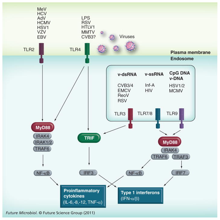
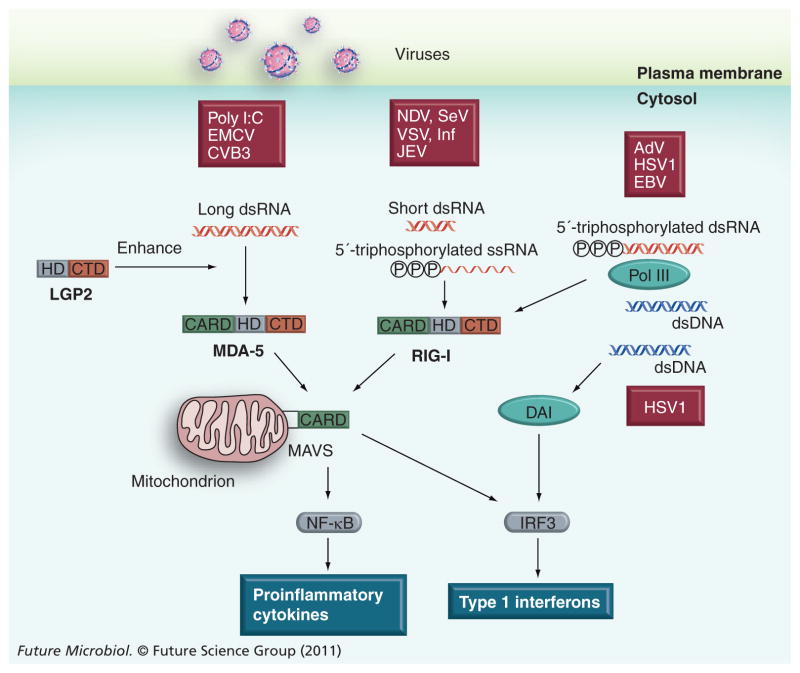
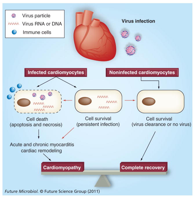
Virus infection can inflict significant damage on cardiomyocytes through direct injury and secondary immune reactions, leading to myocarditis and dilated cardiomyopathy. While viral myocarditis or cardiomyopathy is a complication of systemic infection of cardiotropic viruses, most individuals infected with the viruses do not develop significant cardiac disease. However, some individuals proceed to develop severe virus-mediated heart disease. Recent studies have shown that viral infection of cardiomyocytes is required for the development of myocarditis and subsequent cardiomyopathy. This suggests that viral infection of cardiomyocytes can be an important step that determines the pathogenesis of viral myocarditis during systemic infection. Accordingly, this article focuses on potential defense mechanisms within the cardiomyocyte against virus infection. Understanding of the cardiomyocyte defense against invading viruses may give us novel insights into the pathophysiology of viral myocarditis, and enable us to develop innovative strategies of diagnosis and treatment for this challenging clinical entity.


Figures

Figure 1
Figure 1. Toll-like receptors and their signaling against virus infection
While TLR2 and 4 recognize viral pathogens on the plasma membrane, TLR3, 7/8 and 9 recognize viral RNA or DNA in the endosome. For the induction of type I IFNs, there are two major pathways downstream of TLRs, MyD88-dependent and TRIF-dependent pathways. Based on data from animal models, the TLR3–TRIF pathway seems to play an important role against CVB3 myocarditis. Further study will be necessary for other cardiotropic virus-mediated myocarditis. Moreover, the significance of each TLR signaling pathway in the cardiomyocyte remains to be elucidated. AdV: Adenovirus; EBV: Epstein–Barr virus; HCMV: Human cytomegalovirus; HSV: Herpes simplex virus; HTLV: Human T-lymphotropic virus; Inf-A: Influenza virus A; IRF: Interferon regulatory factor; LPS: Lipopolysaccharide; MCMV: Mouse cytomegalovirus; MeV: Measles virus; ReoV: Reovirus; RSV: Human respiratory syncytial virus; VZV: Varicella zoster virus.
Figure 2
Figure 2. Recently discovered cytosolic viral RNA and DNA sensors and their substrates
The recognition of viral RNA or DNA in the host cytosol activates downstream signaling, resulting in the production of type I IFNs and proinflammatory cytokines to limit viral replication and propagation. While the importance of the MDA-5–MAVS pathway against systemic CVB3 infection has been confirmed, the impact of the pathway on CVB3 infection of the heart and subsequent myocarditis is not fully understood. AdV: Adenovirus; CBV B3: Coxsackievirus B3; CTD: C-terminal domain; DAI: DNA-dependent activator of interferon regulatory factors; EBV: Epstein–Barr virus; EMCV: Encephalomyocarditis virus; HD: DEAD box helicase-like domain; HSV: Herpes simplex virus; JEV: Japanese encephalitis virus; NDV: Newcastle disease virus; RIG-I: Retinoic acid-induced protein-I; SeV: Sendai virus; VSV: Vesicular stomatitis virus.
Figure 3
Figure 3. Potential fate of cardiomyocytes after virus infection of the heart
Infected cardiomyocytes are thought to die due to the direct effect of the infecting virus (direct injury or apoptosis) and/or the attack of immune cells such as cytotoxic T lymphocytes (necrosis or inflammation), which is usually associated with acute and chronic inflammation in the heart. The loss of functional cardiomyocytes can directly affect cardiac function or induce secondary cardiac remodeling, leading to dilated cardiomyopathy in the long run. It is also known that persistence of virus RNA can be observed in cardiomyocytes in the chronic phase of infection. However, it is still controversial whether this persistence can induce cardiomyocyte death or chronic inflammation, or affect disease progress. Interestingly, while the persistence of virus RNA in the heart is clear, the infectious particles have never been isolated from patient samples in the chronic phase of infection to date. In addition, it has not been determined whether cardiomyocytes can remove such persistently infecting virus RNA from the cell. If possible, elucidation of these mechanisms may provide novel insights into diagnostic and therapeutic strategies against viral myocarditis and subsequent cardiomyopathy. Processes that have been or not been well accepted are shown with black or red arrows, respectively.

References

    1. Blauwet LA, Cooper LT. Myocarditis. Prog Cardiovasc Dis. 2010;52(4):274–288. - PMC - PubMed
    1. Rose NR. Myocarditis: infection versus autoimmunity. J Clin Immunol. 2009;29(6):730–737. - PubMed
    1. Cooper LT., Jr Myocarditis. N Engl J Med. 2009;360(15):1526–1538. Comprehensive review of myocarditis from the viewpoint of clinicians. - PMC - PubMed
    1. Dennert R, Crijns HJ, Heymans S. Acute viral myocarditis. Eur Heart J. 2008;29(17):2073–2082. - PMC - PubMed
    1. Baughman KL. Diagnosis of myocarditis: death of Dallas criteria. Circulation. 2006;113(4):593–595. - PubMed

Publication types

MeSH terms