. 2011 Nov;164(5):1445-59.
doi: 10.1111/j.1476-5381.2011.01487.x.

Sinomenine protects against ischaemic brain injury: involvement of co-inhibition of acid-sensing ion channel 1a and L-type calcium channels

Affiliations

Sinomenine protects against ischaemic brain injury: involvement of co-inhibition of acid-sensing ion channel 1a and L-type calcium channels

Wen-Ning Wu et al. Br J Pharmacol. 2011 Nov.

Abstract

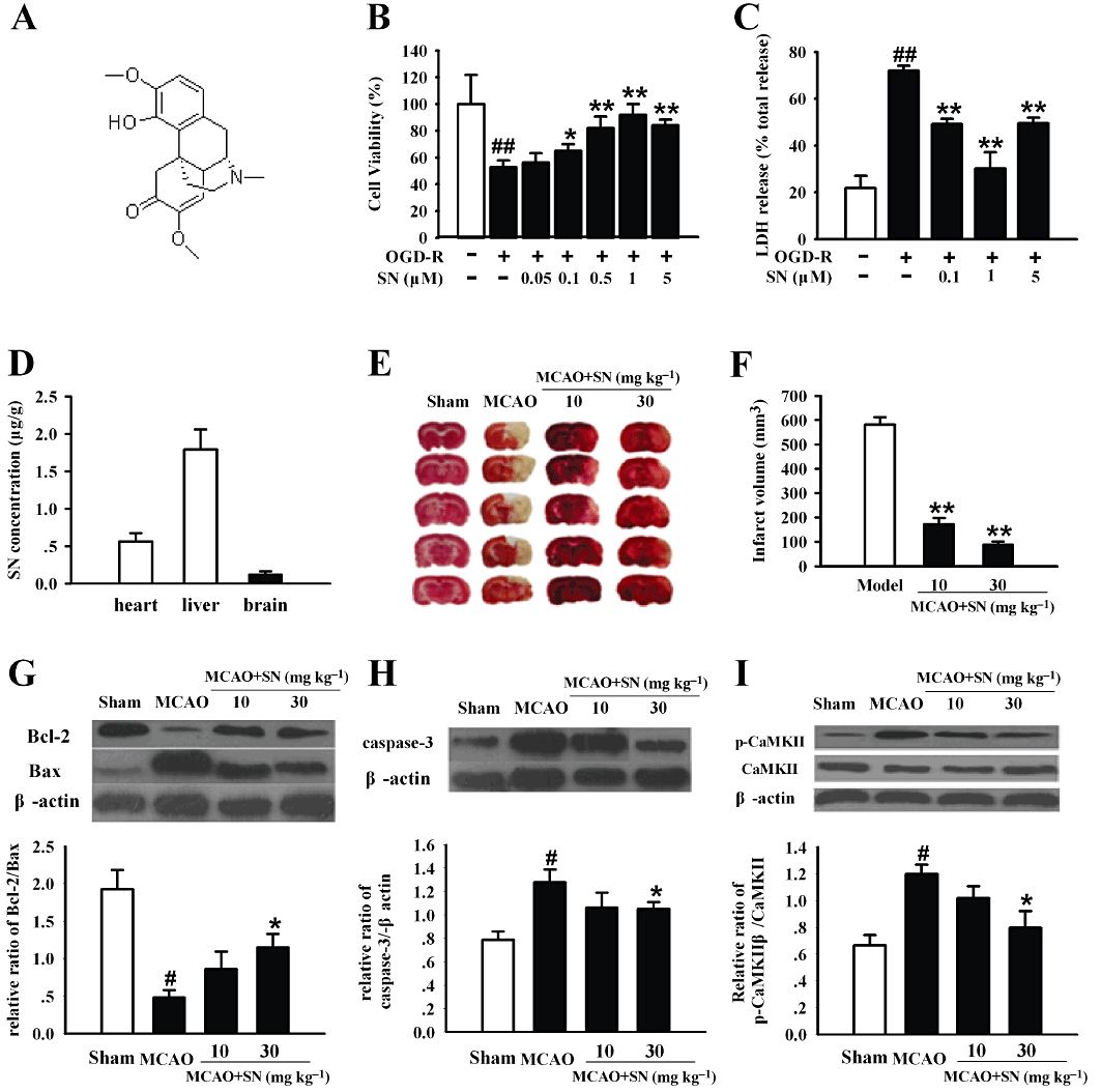
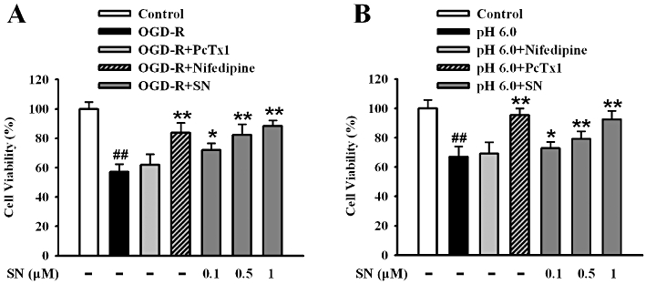
Background and purpose: Sinomenine (SN), a bioactive alkaloid, has been utilized clinically to treat rheumatoid arthritis in China. Our preliminary experiments indicated that it could protect PC12 cells from oxygen-glucose deprivation-reperfusion (OGD-R), we thus investigated the possible effects of SN on cerebral ischaemia and the related mechanism.

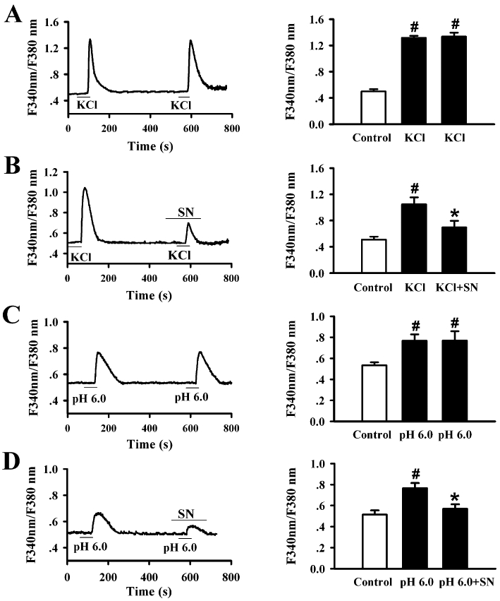
Experimental approach: Middle cerebral artery occlusion in rats was used as an animal model of ischaemic stroke in vivo. The mechanisms of the effects of SN were investigated in vitro using whole-cell patch-clamp recording, calcium imaging in PC12 cells and rat cortical neurons subjected to OGD-R.

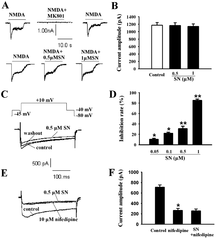
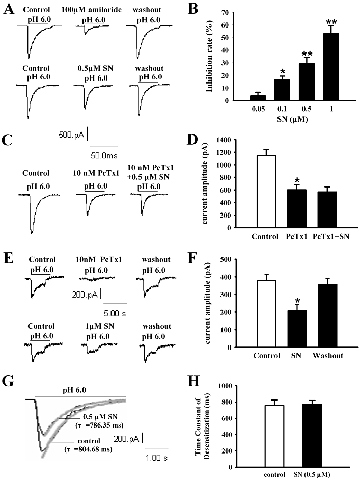
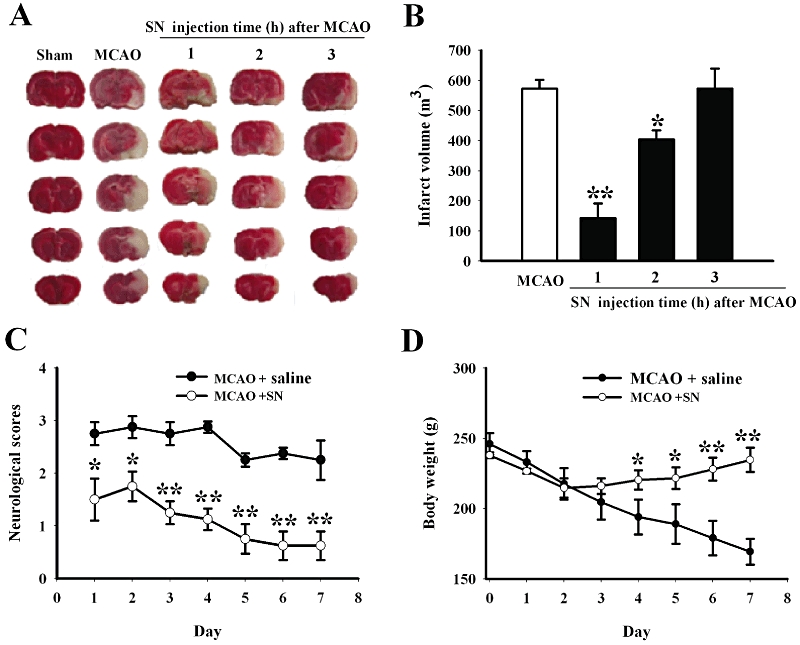
Key results: Pretreatment with SN (10 and 30 mg·kg(-1) , i.p.) significantly decreased brain infarction and the overactivation of calcium-mediated events in rats subjected to 2 h ischaemia followed by 24 h reperfusion. Extracellular application of SN inhibited the currents mediated by acid-sensing ion channel 1a and L-type voltage-gated calcium channels, in the rat cultured neurons, in a concentration-dependent manner. These inhibitory effects contribute to the neuroprotection of SN against OGD-R and extracellular acidosis-induced cytotoxicity. More importantly, administration of SN (30 mg·kg(-1) , i.p.) at 1 and 2 h after cerebral ischaemia also decreased brain infarction and improved functional recovery.

Conclusion and implications: SN exerts potent protective effects against ischaemic brain injury when administered before ischaemia or even after the injury. The inhibitory effects of SN on acid-sensing ion channel 1a and L-type calcium channels are involved in this neuroprotection.


Figures

Figure 1
Figure 1
Pretreatment with SN exhibits protective effects against cerebral ischaemia in vitro and in vivo. (A) The chemical structure of SN. (B) SN reduced OGD-R-induced PC12 cell death in a dose-dependent manner. (C) Different concentrations of SN reduced OGD-R-induced LDH release in PC12 cells. Data are expressed as means ± SEM. n = 6, ##P < 0.01 vs. control,*P < 0.05 and **P < 0.01 vs. OGD-R. (D) SN content was measured using LC-MS in heart, liver and brain at 0.5 h after i.p. injection of 10 mg·kg−1 SN. n = 3. (E) SN attenuated MCAO-induced infarction assessed by TTC staining. The white brain area represents infarcted tissue and the serial sections of each group are from one animal. (F) Infarct volume was measured in the whole brain. Data are expressed as means ± SEM. n = 6. **P < 0.01 vs. MCAO. (G) Representative immunoreactive bands and histogram showing SN attenuated MCAO-induced decrease in the ratio of Bcl-2/Bax. (H) Representative immunoreactive bands showing SN attenuated MCAO-induced increase in activated caspase-3 and histogram representing the quantitative analysis of activated caspase-3 level normalized to β-actin protein. (I) Representative immunoreactive bands showing SN alleviated MCAO-induced up-regulation of CaMKII autophosphorylation and histogram representing the quantitative analysis of autophosphorylated CaMKII level normalized to total protein level. Data are expressed as means ± SEM. n = 3, #P < 0.05 vs. sham and *P < 0.05 vs. MCAO.
Figure 2
Figure 2
SN inhibits VGCCs, but not NMDA currents in rat cultured cortical neurons. SN failed to affect NMDA-activated currents in rat cultured cortical neurons. Representative traces (A) and histogram (B) showing that 0.5 and 1 µM SN failed to inhibit NMDA-activated currents in rat cultured cortical neurons. Data are expressed as means ± SEM and were analysed by Student's paired t-test. n = 4. (C) Representative traces and (D) histogram showing that 0.5 µM SN inhibited IHVA in cortical neurons. (E) and (F) Representative traces and histogram showing that 0.5 µM SN failed to inhibit IHVA in the presence of nifedipine (10 µM) in cultured cortical neurons. Data are expressed as means ± SEM and were analysed by Student's paired t-test. n = 6, *P < 0.05 and **P < 0.01 vs. control.
Figure 3
Figure 3
SN inhibits ASICs in rat cultured cortical neurons and transfected CHO cells. (A) and (B) Representative traces and histogram of results showing that different concentrations of SN inhibited ASICs currents in cortical neurons (n = 6). (C) and (D) Representative traces and histogram of results showing that 0.5 µM SN failed to inhibit ASICs currents in the presence of an ASIC1a blocker in cortical neurons (n = 4). (E) and (F) Representative traces and histogram showing that 10 nM PcTx1 and 1 µM SN inhibited the ASIC1a current expressed in CHO cells (n = 6). Data are expressed as means ± SEM and were analysed by Student's paired t-test, *P < 0.05 and **P < 0.01 vs. control. (G) and (H) Representative traces and summary data showing SN had no effect on the desensitization of ASICs currents in cortical neurons. n = 6.
Figure 6
Figure 6
Administration of SN after cerebral ischaemia protects against brain injury and improves functional recovery. (A) Effect of SN (30 mg·kg−1, i.p) administration, at 1, 2 and 3 h after cerebral ischaemia, on infarct volume induced by 120 min of MCAO, evaluated 24 h after induction of ischaemia. The white brain area represents infarcted tissue. (B) Infarct volume was measured in total brain. Data are expressed as means ± SEM, n = 5. *P < 0.05 and **P < 0.01 vs. MCAO. (C) Neurological scores were graded. SN (30 mg·kg−1) or saline (control) was injected 1 h after cerebral ischaemia and once daily for a week. Data are expressed as means ± SEM. n = 4, *P < 0.05 and **P < 0.01 vs. saline at the same day. (D) Body weights were monitored after administration of saline or SN (30 mg·kg−1) at 1 h after cerebral ischaemia and once daily for a week. Data are expressed as means ± SEM. n = 5, *P < 0.05 and **P < 0.01 vs. saline at the same day.
Figure 4
Figure 4
SN inhibits [Ca2+]i increase induced by KCl and extracellular acidosis in rat cultured cortical neurons. (A) Representative traces and histogram of results showing that [Ca2+]i responds to repeated KCl stimulation under control conditions with a time interval of 5 min between stimuli. Repeated stimulation with KCl (30 mM) produced an equal transient [Ca2+]i increase. n = 7. (B) Representative traces and histogram of results showing that SN inhibited [Ca2+]i increase induced by KCl. n = 10. (C) Representative traces and histogram of results showing that [Ca2+]i responds to repeated acid stimulation (extracellular acid solution, pH = 6.0) under control conditions with a time interval of 5 min between stimuli. n = 8. (D) Representative traces and histogram of results showing that SN inhibited [Ca2+]i increase induced by extracellular acid solution. n = 9. Data are expressed as means ± SEM and were analysed by Student's paired t-test, #P < 0.05 vs. control, *P < 0.05 vs. KCl or pH 6.0.
Figure 5
Figure 5
Antagonism of L-type channels and ASIC1a contribute to the neuroprotection of SN against ischaemic insults in cortical neurons. (A) Nifedipine (10 µM) and different concentrations of SN reduced OGD-R-induced cell death in cultured cortical neurons. (B) PcTx1 and different concentrations of SN reduced extracellular acidosis-induced cell death in cultured cortical neurons. Data are expressed as means ± SEM. n = 8, ##P < 0.01 vs. control, *P < 0.05 and **P < 0.01 vs. OGD-R or pH 6.0.

Similar articles

Cited by

References

    1. Bano D, Nicotera P. Ca2+ signals and neuronal death in brain ischemia. Stroke. 2007;38(Suppl.):674–676. - PubMed
    1. Bao GH, Qin GW, Wang R, Tang XC. Morphinane alkaloids with cell protective effects from Sinomenium acutum. J Nat Prod. 2005;68:1128–1130. - PubMed
    1. Baron A, Waldmann R, Lazdunski M. ASIC-like, proton-activated currents in rat hippocampal neurons. J Physiol. 2002;539:485–494. - PMC - PubMed
    1. Berliocchi L, Bano D, Nicotera P. Ca2+ signals and death programmes in neurons. Philos Trans R Soc Lond B Biol Sci. 2005;360:2255–2258. - PMC - PubMed
    1. Cai F, Li CR, Wu JL, Chen JG, Liu C, Min Q, et al. Theaflavin ameliorates cerebral ischemia-reperfusion injury in rats through its anti-inflammatory effect and modulation of STAT-1. Mediators Inflamm. 2006;2006:30490–30498. - PMC - PubMed

Publication types

MeSH terms