. 2011 May 17:12:21.
doi: 10.1186/1471-2091-12-21.

Mapping of the minimal inorganic phosphate transporting unit of human PiT2 suggests a structure universal to PiT-related proteins from all kingdoms of life

Affiliations

Mapping of the minimal inorganic phosphate transporting unit of human PiT2 suggests a structure universal to PiT-related proteins from all kingdoms of life

Pernille Bøttger et al. BMC Biochem. .

Abstract

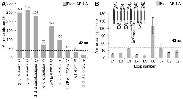
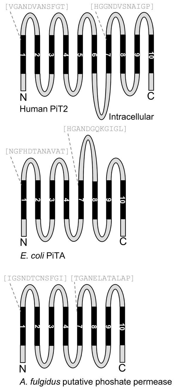
Background: The inorganic (Pi) phosphate transporter (PiT) family comprises known and putative Na(+)- or H(+)-dependent Pi-transporting proteins with representatives from all kingdoms. The mammalian members are placed in the outer cell membranes and suggested to supply cells with Pi to maintain house-keeping functions. Alignment of protein sequences representing PiT family members from all kingdoms reveals the presence of conserved amino acids and that bacterial phosphate permeases and putative phosphate permeases from archaea lack substantial parts of the protein sequence when compared to the mammalian PiT family members. Besides being Na(+)-dependent P(i) (NaP(i)) transporters, the mammalian PiT paralogs, PiT1 and PiT2, also are receptors for gamma-retroviruses. We have here exploited the dual-function of PiT1 and PiT2 to study the structure-function relationship of PiT proteins.

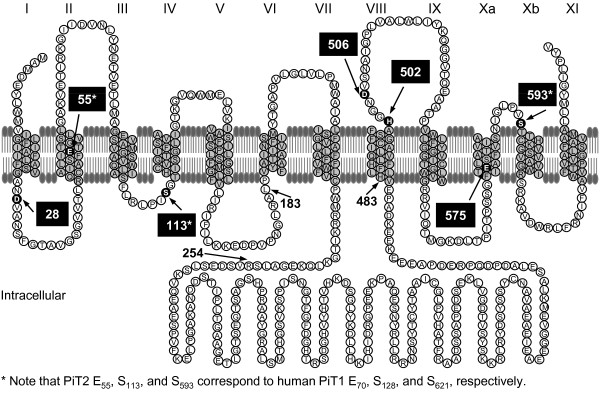
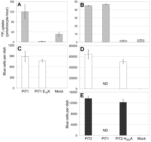
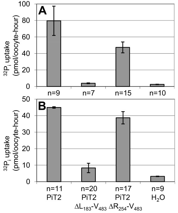
Results: We show that the human PiT2 histidine, H(502), and the human PiT1 glutamate, E(70),--both conserved in eukaryotic PiT family members--are critical for P(i) transport function. Noticeably, human PiT2 H(502) is located in the C-terminal PiT family signature sequence, and human PiT1 E(70) is located in ProDom domains characteristic for all PiT family members.A human PiT2 truncation mutant, which consists of the predicted 10 transmembrane (TM) domain backbone without a large intracellular domain (human PiT2ΔR(254)-V(483)), was found to be a fully functional P(i) transporter. Further truncation of the human PiT2 protein by additional removal of two predicted TM domains together with the large intracellular domain created a mutant that resembles a bacterial phosphate permease and an archaeal putative phosphate permease. This human PiT2 truncation mutant (human PiT2ΔL(183)-V(483)) did also support P(i) transport albeit at very low levels.

Conclusions: The results suggest that the overall structure of the P(i)-transporting unit of the PiT family proteins has remained unchanged during evolution. Moreover, in combination, our studies of the gene structure of the human PiT1 and PiT2 genes (SLC20A1 and SLC20A2, respectively) and alignment of protein sequences of PiT family members from all kingdoms, along with the studies of the dual functions of the human PiT paralogs show that these proteins are excellent as models for studying the evolution of a protein's structure-function relationship.


Figures

Figure 1
Figure 1
Putative topological model of human PiT2 and mutants. Putative membrane topology model of human PiT2 on which the mutant proteins investigated in the present paper are based; the model was originally proposed by O'Hara and coworkers [8,20]. The numbers of the TMs are indicated above the model. Other membrane topology models have been proposed for PiT1 [22,23] and PiT2 [24], which suggested diverging topology for the two paralogs; the alternative PiT2 model is shown in Figure 2. The amino acids previously identified in human PiT2 as being critical for Pi transport function are highlighted with black filling and pointed out with arrows; D28, E55, S113, D506, E575, and S593[18,27,28]. In human PiT1, the amino acids S128 (PiT2 S113) and S621 (PiT2 S593) have previously been identified as being critical for PiT1 Pi transport function [29]. In the present study, human PiT2 H502 (situated in the PiT family signature sequence) and human PiT1 E70 (equivalent in position to human PiT2 E55) are also identified as critical for Pi transport function (see Figure 3). The grey-filled sequences (L11-L161 and V492-V640), represent the N- and C-terminal, respectively, ProDom domains (PD001131) published in 2004 defining the PiT family members [27]. The dark grey-filed sequence (I53-L127) represents the most recent ProDom domain defining the PiT family members http://prodom.prabi.fr/.
Figure 2
Figure 2
Alternative topological model of human PiT2 and mutants. Membrane topology model for human PiT2 suggested by Salaün and coworkers [24]; the TMs are shown as grey-filled sequences and their numbers are indicated with roman numbers above the model. This model shares some similarity to a membrane topology model for PiT1 proposed in 2002 [22]. Based on the cellular location of C-terminal tags, the C-terminal ends of PiT1 and PiT2 were predicted to be extracellular [22,24]. And based on the cellular location of an N-terminal tag on PiT2 and glycosylation of a site in human PiT1 and partly glycosylation of the same site in human PiT2 although oddly not in hamster PiT2, the N-termini of PiT1 and PiT2 were suggested to be extracellular [22,24]; due to a suggested additional TM after TM3 in Figure 1 (TMIV in this figure), this did not influence the orientation of the large intracellular domain in these models compared to the model in Figure 1. The PiT2 model shown in Figure 1 and this figure, respectively, and the PiT1 model proposed in 2002 [22] were later compared by us [18]. In 2009, Farrell and coworkers proposed a modified model of human PiT1 based on substituted cysteine accessibility mutagenesis [23]. The recent model of PiT1 shows more resemblance to the PiT2 models shown in this figure and in Figure 1 concerning the length and position of the large intracellular domain (L6) than the model from 2002. The amino acids identified in human PiT2 and human PiT1 as being critical for Pi transport function are highlighted with black filling and pointed out with arrows; for references see legend to Figure 1. Compared to the PiT2 model in Figure 1, the PiT2 model proposed by Salaün and coworkers (this figure) and the PiT1 model proposed in 2009 by Farrell and coworkers do not affect the placement of PiT1 D43 (PiT2 D28), PiT1 E70 (PiT2 E55), PiT1 H530 (PiT2 H502), PiT1 D534 (PiT2 D506), PiT1 E603 (PiT2 E575), and PiT1 S621 (PiT2 S593) in either a TM domain or a loop sequence [23,24] (compare Figure 1 and this figure). However, PiT1 S128 (PiT2 S113) placed in loop regions in the PiT2 models (this figure and Figure 1), is in the PiT1 model from 2009 suggested to be placed in a TM domain [23].
Figure 3
Figure 3
Analysis of human PiT1 E70K and PiT2 H502A for Na32Pi uptake and gamma-retroviral receptor function. A-B X. laevis oocytes were injected with H2O (Mock) or cRNA of the indicated constructs. Three days later, a 32Pi uptake assay was performed and the 32Pi uptake in individual oocytes was measured. Data are the mean value of (n) numbers of oocytes ±SEM, see Additional File 2 for data and statistics. Experiments A and B were made independently of each other, and the experiments were repeated and similar results obtained. C CHO K1 cells were transfected with CsCl-purified PiT1- or PiT1 E70K-encoding plasmid or empty vector DNA (Mock). Three independent precipitates were made for each construct. Forty-eight hours after transfection, approx. 8 × 10⁴ 10A1 MLV pseudotypes were added per dish. The average numbers (±SEM) of blue (infected) cells per dish from three dishes receiving independent precipitates are shown, see Additional File 2 for data and statistics. D-E were made in parallel using the same protocol as in (C) with the exception that Nucleobond-purified plasmids encoding PiT2, PiT2 H502A, or empty vector DNA were used. The dishes were challenged with approx. 4 × 10⁴ 10A1 MLV pseudotypes (D) or A-MLV pseudotypes (E). The average numbers (±SEM) of blue (infected) cells per dish from three dishes receiving independent precipitates are shown, see Additional File 2 for data and statistics.
Figure 4
Figure 4
Investigation of the loop sequence length in PiT family members. A The amino acid lengths of loop 6 (L6) are plotted for nine PiT family members (H. sapiens PiT2, H. sapiens PiT1, N. crassa Pho-4+, A. thaliana Pht2_1, E. coli PiTA, and putative phosphate permeases from D. melanogaster, C. elegans, T. brucei, and A. fulgidus). The L6 lengths are defined by the predicted TM domains in the protein sequences of the PiT family members; see alignment in and legend to Additional File 1 Figure A (AF 1 A). The maximum limit of a loop length (42 amino acids) estimated in Figure 4B is indicated on the figure. It illustrates that loop lengths at 1 to 42 amino acids define a loop sequence and loop lengths at 43 amino acids or higher defines a domain. B The numbers of amino acids in loop 1 (L1) to loop 9 (L9) in the protein sequences listed in the legend to A are shown. The loop lengths were defined by the sequences connecting the predicted TM domains in the protein sequences for the nine PiT family members; see alignment in and legend to Additional File 1 Figure A (AF 1 A). Data are the mean value of (n) numbers of loops counted ±SEM, see Additional File 2 for data. The stippled line indicates the maximum length for a loop sequence (L3) which is ~ 42 amino acids given with 95% confidence (38.6 ±3.4 amino acids ~ 35 to 42 amino acids). Note that the 95% confidence interval for L7 is 42.9 ±28.8 amino acids, illustrating that this loop length is subjected to high uncertainty because of an unusually long L7 in E. coli PiTA. The 95% confidence interval for L7 calculated when excluding L7 E. coli PiTA is 28.3 ±2.4 amino acids. The topology model indicates the positions of L1 to L9; stippled loops indicate the observed variable lengths of L6 (the large intracellular domain).
Figure 5
Figure 5
Predicted topologies of H. sapiens PiT2, E. coli PiTA, and A. fulgidus putative phosphate permease. Illustrations of the putative topology of H. sapiens PiT2, E. coli PiTA, and A. fulgidus putative phosphate permease are shown. TM domains were predicted using the TMHMM server (H. sapiens PiT2) and the DAS server (E. coli PiTA and A. fulgidus putative phosphate permease) (Additional File 1 Figure A), see "Methods" for description. The N-terminal and C-terminal PiT-family signature sequences [18] are given in grey letters, and grey stippled lines indicate the predicted placement.
Figure 6
Figure 6
Na32Pi uptake mediated by human PiT2 and truncation mutants analyzed in X. laevis oocytes. Oocytes were injected with H2O or cRNA of the indicated constructs. Two (experiment A) or three (experiment B) days later, a 32Pi uptake assay was performed and the 32Pi uptake in individual oocytes was measured. Data are the mean value of (n) numbers of oocytes ±SEM, see Additional File 2 for data and statistics.

References

    1. Oláh Z, Lehel C, Anderson WB, Eiden MV, Wilson CA. The cellular receptor for gibbon ape leukemia virus is a novel high affinity sodium-dependent phosphate transporter. J Biol Chem. 1994;269(41):25426–25431. - PubMed
    1. Kavanaugh MP, Miller DG, Zhang W, Law W, Kozak SL, Kabat D, Miller AD. Cell-surface receptors for gibbon ape leukemia virus and amphotropic murine retrovirus are inducible sodium-dependent phosphate symporters. Proc Natl Acad Sci USA. 1994;91(15):7071–7075. doi: 10.1073/pnas.91.15.7071. - DOI - PMC - PubMed
    1. Kavanaugh MP, Kabat D. Identification and characterization of a widely expressed phosphate transporter/retrovirus receptor family. Kidney Int. 1996;49(4):959–963. doi: 10.1038/ki.1996.135. - DOI - PubMed
    1. Virkki LV, Biber J, Murer H, Forster IC. Phosphate transporters: a tale of two solute carrier families. Am J Physiol Renal Physiol. 2007;293(3):F643–654. doi: 10.1152/ajprenal.00228.2007. - DOI - PubMed
    1. Wain HM, Bruford EA, Lovering RC, Lush MJ, Wright MW, Povey S. Guidelines for human gene nomenclature. Genomics. 2002;79(4):464–470. doi: 10.1006/geno.2002.6748. - DOI - PubMed

Publication types

MeSH terms

Substances