. 2011 Jul 15;286(28):25076-87.
doi: 10.1074/jbc.M110.205328. Epub 2011 May 17.

Peptide-protein interactions suggest that acetylation of lysines 381 and 382 of p53 is important for positive coactivator 4-p53 interaction

Affiliations

Peptide-protein interactions suggest that acetylation of lysines 381 and 382 of p53 is important for positive coactivator 4-p53 interaction

Subrata Debnath et al. J Biol Chem. .

Abstract

The human transcriptional positive coactivator 4 (PC4) activates several p53-dependent genes. It has been demonstrated that this is a consequence of direct interaction with p53. Previously, we have concluded that PC4 interacts mainly with the C-terminal negative regulatory domain of p53 through its DNA binding C-terminal half. NMR chemical shift perturbation studies with peptide fragments indicated that amino acids 380-386 of p53 are crucial for interaction with PC4. This was verified by fluorescence anisotropy and sedimentation velocity studies. A peptide consisting of p53-(380-386) sequence, when attached to a cell penetration tag and nuclear localization signal, localizes to the nucleus and inhibits luciferase gene expression from a transfected plasmid carrying a Luc gene under a p53-dependent promoter. Acetylation of lysine 382/381 enhanced the binding of this peptide to PC4 by about an order of magnitude. NMR and mutagenesis studies indicated that serine 73 of PC4 is an important residue for recognition of p53. Intermolecular nuclear Overhauser effect placed aspartate 76 in the vicinity of lysine 381, indicating that the region around residues 73-76 of PC4 is important for p53 recognition. We conclude that the 380-386 region of p53 interacts with the region around residues 73-76 of PC4, and acetylation of lysine 382/381 of p53 may play an important role in modulating p53-PC4 interaction and as a consequence PC4 mediated activation of p53 target genes.


Figures
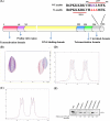
FIGURE 1.
FIGURE 1.
A, shown is the domain structure of p53. B, overlap of the methyl region TOCSY spectra of p53-(364–393) alone (blue) and p53-(364–393) in the presence of PC4 (10:1) (red) is shown. C, overlap of the one-dimensional spectra of the methyl region of p53-(366–372) alone (blue) and p53-(366–372) in the presence of PC4 (7:1) (red) is shown. D, overlap of the one-dimensional spectra of the methyl region of p53-(380–386) alone (blue) and p53-(380–386) in the presence of PC4 (7:1) (red) is shown. All the experiments were conducted at 37 °C on a 600-MHz Bruker Biospin NMR spectrometer using TCI cryoprobe. The buffer was 50 mm potassium phosphate buffer, pH 6.2, containing 100 mm KCl. TOCSY (mixing time 60 ms) spectra were recorded with solvent suppression, which was achieved using water suppression by gradient-tailored excitation, and the spinlock in TOCSY experiment was attained by the MLEV sequence. E, shown is a GST pulldown assay. 1 μg of GST (lane 2) or GST-p53 point mutant proteins (third to seventh lanes) were incubated with bacterial lysate containing 200 ng of PC4 and analyzed by immunoblotting with anti-PC4 antibody. First lane, 20% input of bacterial cell lysate is shown. Intensities relative to the GST only control are reported at the bottom.
FIGURE 2.
FIGURE 2.
Sedimentation velocity run of fluorescein end-labeled p53-(380–386). A, p53-(380–386) alone (SEDPHAT profile) is shown. B, sedimentation velocity run of fluorescein end-labeled p53-(380–386) complex with PC4 (1:1) is shown. The scan was recorded at 495 nm. C, shown is a binding isotherm of fluorescein end-labeled p53-(380–386) with PC4 (solid diamond) and binding isotherm of fluorescein end-labeled p53-(380–386-K381A/K382A/L383A) with PC4 (circles). D, shown is the effect of unlabeled p53-(380–386) (open circles) and p53-(380–386-K381A/K382A/L383A) (solid circles) on fluorescein-labeled p53-(380–386)·PC4 complex. Fluorescence anisotropy measurements were performed at 4 °C by using a Quantamaster 6 (PTI) T-geometry fluorometer. The titrations were carried out in 20 mm Tris-HCl buffer, pH 7.4, containing 100 mm KCl, 0.2 mm EDTA, 20% glycerol, and 0.1% Nonidet P-40. Fluorescence anisotropy was measured with excitation at 490 nm and emission at 530 nm using bandwidths 5 nm. Further details are given under “Experimental Procedures.”
FIGURE 3.
FIGURE 3.
Binding isotherms of acetylated p53 peptides. Shown is a binding isotherm of end-labeled p53-(380–386-K381Ac) (red), end-labeled p53-(380–386-K382Ac) (green), end-labeled p53-(380–386-K386Ac), and end-labeled p53-(380–386-K381Ac/K382Ac/K386Ac) (black) with PC4. Fluorescence anisotropy measurements were performed at 4 °C by using Quantamaster 6 (PTI) T-geometry fluorometer. The titrations were carried out in 20 mm Tris-HCl buffer, pH 7.4, containing 100 mm KCl, 0.2 mm EDTA, 20% glycerol, and 0.1% Nonidet P-40. Fluorescence anisotropy was measured with excitation at 490 nm and emission at 530 nm using bandwidths 5 nm. The anisotropic data were fitted into a single site binding equation as described before (27).
FIGURE 4.
FIGURE 4.
Inhibition of PC4 enhancement of p53 activation of gene expression by p53-(380–386). p53−/− H1299 cells were transfected with PG13-Luc (500 ng), CMV-β-gal (500 ng), p53 (200 ng), and PC4 (1.2 μg) in combination as indicated. After 14 h of transfection, WT peptide (WT) or control peptide (3A) were added in the indicated lanes. After 24 h of transfection, the luciferase activity was measured and normalized with β-galactosidase activity. Error bars show the S.E. of the replicates. +, present; −, absent.
FIGURE 5.
FIGURE 5.
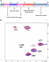
Binding of acetylated p53 peptides by NMR. A, shown is a schematic of the modular structure of PC4. B, binding of PC4 and p53-(380–386) by NMR spectroscopy (1H,15N HSQC) is shown. 1H,15N HSQC overlaps of p53-(380–386-K381A/K382A/L383A)·15N-PC4 (1:1) complex (blue) and p53-(380–386-K381KAc/K382KAc/K386KAc)·15N-PC4 (1:1) complex (red) are shown. NMR experiments were conducted at 37 °C on a 600-MHz Bruker Biospin NMR spectrometer using TCI cryoprobe. The buffer was 50 mm potassium phosphate buffer, pH 6.2, containing 50 mm KCl and 50 mm d5-glycine.
FIGURE 6.
FIGURE 6.
Interaction of p53-(380–386) with mutant PC4. A, shown is a GST pulldown assay of p53 and mutant PC4 interaction; 1 μg of GST-p53 protein was incubated individually with bacterial lysate containing 200 ng of either PC4WT or PC4S73A, pulled down as described under “Experimental Procedures,” and analyzed by immunoblotting with anti-PC4 antibody. Lanes 1 and 2 depict 20% input of bacterial cell lysate containing PC4WT and PC4S73A, respectively. B, 1 μg of GST-p53 protein was incubated individually with bacterial lysate containing 200 ng of either PC4WT or PC4Q65A, pulled down, and analyzed by immunoblotting with anti-PC4 antibody. Lanes 1 and 2 depict 20% input of bacterial cell lysate containing PC4WT and PC4Q65A respectively. C, shown is a binding isotherm of fluorescein end-labeled p53-(380–386) with PC4 S73A. Fluorescence anisotropy measurements were performed at 4 °C by using Quantamaster 6 (PTI) T-geometry fluorometer. The titrations were carried out in 20 mm Tris-HCl buffer, pH 7.4, containing 100 mm KCl, 0.2 mm EDTA, 20% glycerol, and 0.1% Nonidet P-40. Fluorescence anisotropy was measured with excitation at 490 nm and emission at 530 nm using bandwidths 5 nm. D, shown is gene expression activation by wild-type or mutant PC4; p53−/− H1299 cells were transfected with PG13-Luc (500 ng), CMV-β-gal (500 ng), p53 (200 ng), PC4WT (1.2 μg), PC4Q65A (1.2 μg), or PC4S73A (1.2 μg) in combinations as indicated. After 24 h of transfection, the luciferase activity was measured and normalized with β-galactosidase activity. Error bars show the S.E. of the replicates. +, present; −, absent.
FIGURE 7.
FIGURE 7.
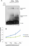
DNA binding by p53 in the presence of PC4. A, 3 ng of a γ-32P-labeled oligonucleotide containing a p53 binding site were incubated with 50 ng of p53 either in the absence of PC4 (lane 2) or with increasing concentrations of PC4 (lanes 3, 4, 5, and 6) or PC4S73A (lanes 7, 8, 9, and 10). Lane 1 contains the γ-32P-labeled oligonucleotide alone. + present; − absent. B, quantitative representation of the effect of PC4 and its point mutant S73A on p53-mediated DNA binding is shown. The levels of induction of p53-mediated binding are represented on the y axis, and the increasing concentrations of PC4 or its point mutant PC4S73A are represented on the x axis. Error bars are obtained from statistical analysis of multiple experiments.
FIGURE 8.
FIGURE 8.
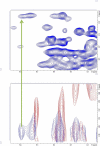
Isotope-edited NOESY spectra. Overlaps of isotope-edited NOESY spectra of p53-(380–386- K381Ac)·PC4 complex (1:1) (blue) and p53(380–386- K381KAc-d3)·PC4 complex (1:1) (red) are shown. The upper spectra show the 1H,15N HSQC of p53-(380–386-K381Ac)-PC4 complex (1:1). Experiments were conducted at 37 °C on a 600-MHz Bruker Biospin NMR spectrometer using TCI cryoprobe. The buffer was 50 mm potassium phosphate buffer, pH 6.2, containing 50 mm KCl and 50 mm d5-glycine.
FIGURE 9.
FIGURE 9.
Depiction of the space-filling model of the C-terminal domain of PC4 (PDB code 2C62). The two subunits are in gray and white. The following residues are highlighted: serine 73 (blue), aspartate 76 (red), aspartate 84 (green), and glutamine 65 (cyan). The bound ssDNA is in a wireframe model.

Similar articles

Cited by

References

    1. Meek D. W., Anderson C. W. (2009) Cold Spring Harb. Perspect. Biol. 1, a000950 - PMC - PubMed
    1. Momand J., Zambetti G. P., Olson D. C., George D., Levine A. J. (1992) Cell 69, 1237–1245 - PubMed
    1. Dornan D., Wertz I., Shimizu H., Arnott D., Frantz G. D., Dowd P., O'Rourke K., Koeppen H., Dixit V. M. (2004) Nature 429, 86–92 - PubMed
    1. Leng R. P., Lin Y., Ma W., Wu H., Lemmers B., Chung S., Parant J. M., Lozano G., Hakem R., Benchimol S. (2003) Cell 112, 779–791 - PubMed
    1. Haupt Y., Maya R., Kazaz A., Oren M. (1997) Nature 387, 296–299 - PubMed

Publication types

MeSH terms