. 2011 Jul 8;286(27):24508-18.
doi: 10.1074/jbc.M111.234062. Epub 2011 May 17.

The Mycobacterium tuberculosis early secreted antigenic target of 6 kDa inhibits T cell interferon-γ production through the p38 mitogen-activated protein kinase pathway

Affiliations

The Mycobacterium tuberculosis early secreted antigenic target of 6 kDa inhibits T cell interferon-γ production through the p38 mitogen-activated protein kinase pathway

Hui Peng et al. J Biol Chem. .

Abstract

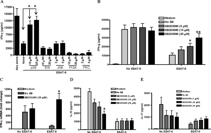
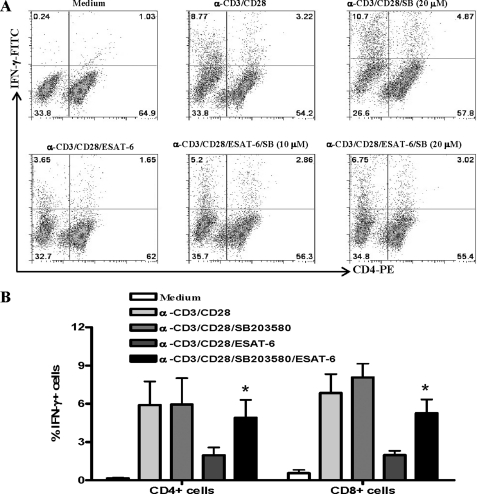
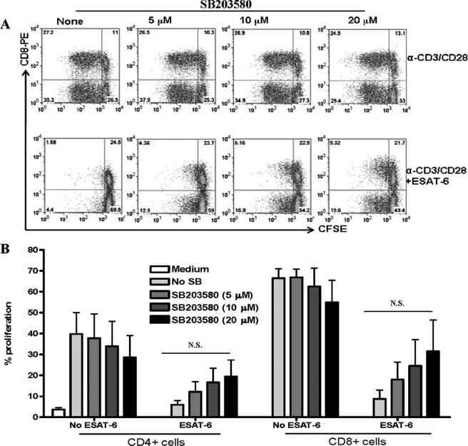
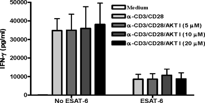
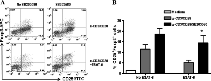
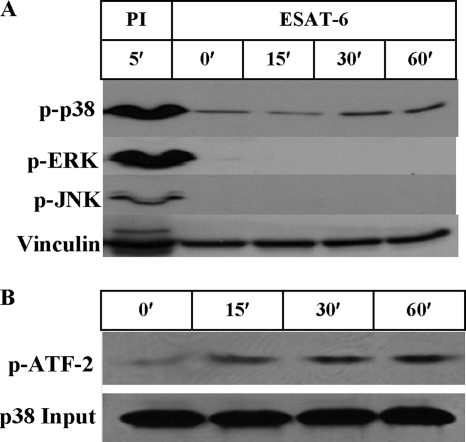
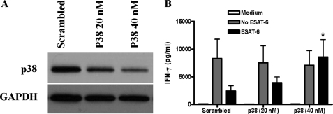
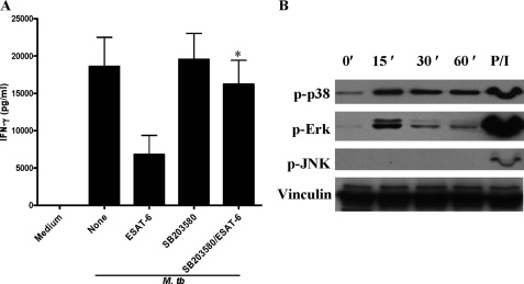
We reported previously that the early secreted antigenic target of 6 kDa (ESAT-6) from Mycobacterium tuberculosis directly inhibits human T cell IFN-γ production and proliferation in response to stimulation with anti-CD3 and anti-CD28. To determine the mechanism of this effect, we treated T cells with kinase inhibitors before stimulation with ESAT-6. Only the p38 MAPK inhibitor, SB203580, abrogated ESAT-6-mediated inhibition of IFN-γ production in a dose-dependent manner. SB203580 did not reverse ESAT-6-mediated inhibition of IL-17 and IL-10 production, suggesting a specific effect of SB203580 on IFN-γ production. SB203580 did not act through inhibition of AKT (PKB) as an AKT inhibitor did not affect ESAT-6 inhibition of T cell IFN-γ production and proliferation. ESAT-6 did not reduce IFN-γ production by expanding FoxP3(+) T regulatory cells. Incubation of T cells with ESAT-6 induced phosphorylation and increased functional p38 MAPK activity, but not activation of ERK or JNK. Incubation of peripheral blood mononuclear cells with ESAT-6 induced activation of p38 MAPK, and inhibition of p38 MAPK with SB203580 reversed ESAT-6 inhibition of M. tuberculosis-stimulated IFN-γ production by peripheral blood mononuclear cells from subjects with latent tuberculosis infection. Silencing of p38α MAPK with siRNA rendered T cells resistant to ESAT-6 inhibition of IFN-γ production. Taken together, our results demonstrate that ESAT-6 inhibits T cell IFN-γ production in a p38 MAPK-dependent manner.


Figures

FIGURE 1.
FIGURE 1.
Effects of PK inhibitors on ESAT-6 inhibition of T cell IFN-γ production. A, purified T cells from five healthy individuals were incubated with medium or different PK inhibitors for 1 h and then cultured with ESAT-6 (3.3 μm) for 1 h. The cells were then plated in a 96-well plate coated with anti-CD3 and anti-CD28 for 48 h, and IFN-γ concentrations in the supernatants were measured by ELISA. IFN-γ levels were negligible in supernatants of unstimulated cells. Means and S.E. are shown. *, p = 0.007, as compared with ESAT-6-treated cells without inhibitors. Abs alone, antibodies alone. B, purified T cells from 13 healthy individuals were treated with medium or SB203580 and then treated with or without ESAT-6 and incubated with anti-CD3 and anti-CD28 for 48 h, as in panel A. IFN-γ levels in the supernatants were measured by ELISA. Means and S.E. are shown. *, p = 0.03, **, p = 0.001, as compared with cells treated with ESAT-6 alone. No SB, no SB203580 inhibitor. C, purified T cells from four donors were treated as described in panel B, total cellular RNA was isolated, and IFN-γ mRNA was measured by real-time PCR, using 18 S rRNA as an internal control. Means and S.E. are shown. *, p < 0.05, as compared with the cells with ESAT-6 without SB203580. D and E, purified T cells from five donors were treated as described in panel B, and the concentrations of IL-10 (D) and IL-17 (E) in the culture supernatants were measured by ELISA. Means and S.E. are shown. *, p < 0.05, as compared with the cells stimulated with anti-CD3 and anti-CD28 with no SB203580 (D).
FIGURE 2.
FIGURE 2.
Effect of inhibition of p38 MAPK on ESAT-6-mediated reduction of IFN-γ+ CD4+ and CD8+ T cells. Purified T cells from healthy subjects were treated with medium or different concentrations of SB203580 for 1 h and then with or without 3.3 μm ESAT-6 for 1 h. The cells were then cultured with plate-bound anti-CD3 and anti-CD28. After 48 h, the cells were collected and stained with anti-CD4 and anti-IFN-γ and analyzed by flow cytometry. A, a representative result is shown. B, means and S.E. for results with five donors are shown. *, p < 0.05, as compared with cells treated with ESAT-6 alone.
FIGURE 3.
FIGURE 3.
Effect of inhibition of p38 MAPK on ESAT-6-mediated reduction of anti-CD3-induced T cell proliferation. Purified T cells from four donors were labeled with CFSE and treated with medium or different concentrations of SB203580 for 1 h and then with or without 3.3 μm ESAT-6 for 1 h. The cells were then cultured with plate-bound anti-CD3 and anti-CD28. After 96 h, the cells were stained with PE-anti-CD8, and cell proliferation was analyzed by CFSE dilution, using flow cytometry. A, a representative result is shown. B, means and S.E. of four different experiments are shown for CD4+ and CD8+ cells. No SB, no SB203580 inhibitor. N.S., not significant.
FIGURE 4.
FIGURE 4.
ESAT-6 inhibition of T cell IFN-γ production is independent of AKT. Purified T cells were treated with medium or different concentrations of AKT inhibitor for 1 h and then with or without 3.3 μm ESAT-6 for 1 h. The cells were then cultured with plate-bound anti-CD3 and anti-CD28 for 48 h. The supernatants were collected, and IFN-γ concentrations were measured by ELISA. Means and S.E. for results with four donors are shown.
FIGURE 5.
FIGURE 5.
The effects of ESAT-6 and SB203580 on IFN-γ production are not mediated by effects on Tregs. Purified human T cells were treated with medium or SB203580 for 1 h and then with or without 3.3 μm ESAT-6 for 1 h. The cells were then cultured with plate-bound anti-CD3 and anti-CD28. After 72 h, the cells were collected, washed, and stained with anti-CD4 and anti-CD25 followed by intracellular staining with anti-Foxp3. After gating on CD4+ cells, the frequency of CD25+Foxp3+ cells (Tregs) was determined. A, a representative result is shown. B, means and S.E. from experiments with four donors are shown. *, p < 0.05, as compared with ESAT-6-treated cells without SB203580.
FIGURE 6.
FIGURE 6.
ESAT-6 directly activates p38 MAPK in T cells. A, purified T cells were incubated with 3.3 μm ESAT-6 for different periods or stimulated with phorbol myristate acetate and ionomycin (PI) for 5 min as a positive control. The cells were collected, and total protein extracts were prepared. Western blotting was performed with phospho-specific (p) antibodies against p38, ERK, and JNK. The same blot was stripped and blotted for vinculin as a protein loading control. A representative result of experiments performed on T cells from four donors is shown. B, T cells were treated with 3.3 μm ESAT-6 for different periods, and total cell protein extracts were prepared. Phosphorylated p38 MAPK was immunoprecipitated with anti-phospho-p38, and the kinase activity in the immunoprecipitates was determined by Western blotting to measure the ability to phosphorylate the p38 MAPK substrate, recombinant ATF-2, as detailed under “Materials and Methods.” Total p38 MAPK levels in the protein extracts before immunoprecipitation, measured by Western blotting, were used as input controls. A representative result from experiments with three donors is shown.
FIGURE 7.
FIGURE 7.
Effect of ESAT-6 on calcium influx in T cells. Purified T cells from healthy donors were loaded with a Ca2+-sensitive fluorescent dye and then cultured in medium alone, with ESAT-6 or with anti-CD3 and anti-CD28 for 1 h at room temperature. The cells were then incubated at 37 °C for 5 min, after which Ca2+ influx was measured for 250 s (x axis) by flow cytometry as the mean fluorescence intensity (MFI) for fluo-4 (y axis). The basal Ca2+ influx in untreated cells is shown as the gray area. A representative result from four experiments is shown.
FIGURE 8.
FIGURE 8.
The effect of silencing p38 MAPK on ESAT-6-mediated inhibition of T cell IFN-γ production. Purified human T cells were transfected with scrambled or p38 MAPK siRNA for 48 h. A, the silencing efficiency of siRNA was evaluated by Western blotting of total T cell protein extracts with anti-p38α MAPK. The membranes were stripped and blotted with anti-GAPDH to control for protein loading. A representative result of four different experiments is shown. B, siRNA-transfected cells were incubated, with or without 3.3 μm ESAT-6, for 1 h before further treatment with plate-bound anti-CD3 and anti-CD28. After 72 h, the supernatants were collected, and IFN-γ levels were measured by ELISA. Means and S.E. from experiments with five donors are shown. *, p < 0.05, as compared with ESAT-6-treated cells with scrambled siRNA.
FIGURE 9.
FIGURE 9.
ESAT-6 inhibits IFN-γ production by M. tuberculosis-stimulated PBMC through activation of p38 MAPK. A, PBMC from seven donors with latent tuberculosis infection were treated with medium or SB203580 (20 μm) for 1 h and then with or without 3.3 μm ESAT-6 for 1 h. The cells were then incubated with heat-killed M. tuberculosis (M. tb). After 48 h, supernatants were collected, and IFN-γ concentrations were measured by ELISA. Means and S.E. are shown. *, p = 0.04, as compared with ESAT-6-treated cells. B, PBMC from three donors with latent tuberculosis infection were incubated with 3.3 μm ESAT-6 for different time periods, total cell protein extracts were prepared, and activation of p38, ERK, and JNK MAPK was evaluated by Western blotting with phospho-specific (p) antibodies. Vinculin was used as a protein loading control. PBMC were stimulated with phorbol myristate acetate and ionomycin (P/I) as a positive control. A representative result is shown.

Similar articles

Cited by

References

    1. Flynn J. L., Chan J. (2001) Annu. Rev. Immunol. 19, 93–129 - PubMed
    1. Mogues T., Goodrich M. E., Ryan L., LaCourse R., North R. J. (2001) J. Exp. Med. 193, 271–280 - PMC - PubMed
    1. Daley C. L. (2010) Am. J. Respir Crit Care Med. 181, 550–555 - PubMed
    1. Jassal M. S., Bishai W. R. (2010) Clin. Infect. Dis. 50, Suppl. 3, S156–S164 - PMC - PubMed
    1. Andersen P., Andersen A. B., Sørensen A. L., Nagai S. (1995) J. Immunol. 154, 3359–3372 - PubMed

Publication types

MeSH terms