. 2011 Sep 1;39(16):7161-78.
doi: 10.1093/nar/gkr234. Epub 2011 May 17.

G4 resolvase 1 tightly binds and unwinds unimolecular G4-DNA

Affiliations

G4 resolvase 1 tightly binds and unwinds unimolecular G4-DNA

Banabihari Giri et al. Nucleic Acids Res. .

Abstract

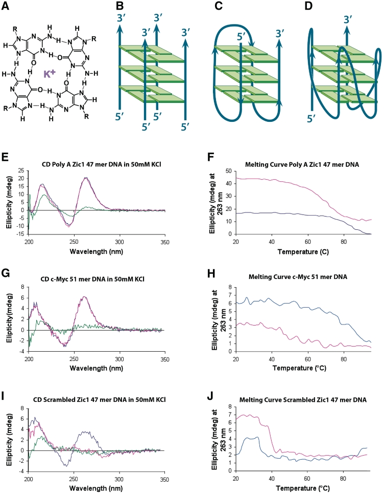
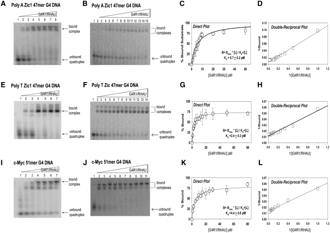
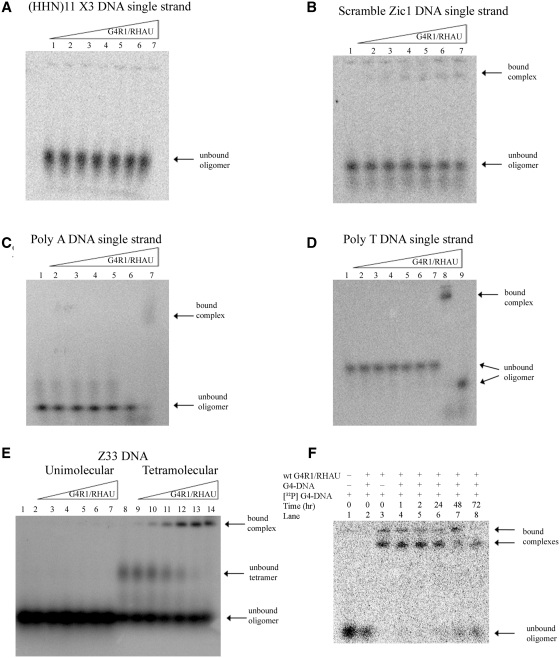
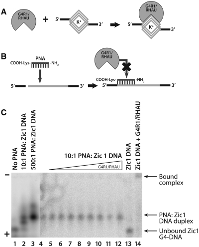
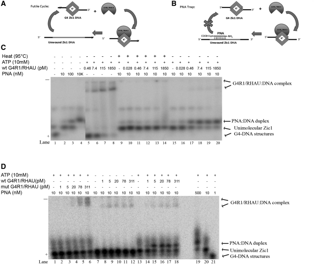
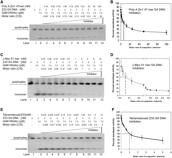
It has been previously shown that the DHX36 gene product, G4R1/RHAU, tightly binds tetramolecular G4-DNA with high affinity and resolves these structures into single strands. Here, we test the ability of G4R1/RHAU to bind and unwind unimolecular G4-DNA. Gel mobility shift assays were used to measure the binding affinity of G4R1/RHAU for unimolecular G4-DNA-formed sequences from the Zic1 gene and the c-Myc promoter. Extremely tight binding produced apparent K(d)'s of 6, 3 and 4 pM for two Zic1 G4-DNAs and a c-Myc G4-DNA, respectively. The low enzyme concentrations required for measuring these K(d)'s limit the precision of their determination to upper boundary estimates. Similar tight binding was not observed in control non-G4 forming DNA sequences or in single-stranded DNA having guanine-rich runs capable of forming tetramolecular G4-DNA. Using a peptide nucleic acid (PNA) trap assay, we show that G4R1/RHAU catalyzes unwinding of unimolecular Zic1 G4-DNA into an unstructured state capable of hybridizing to a complementary PNA. Binding was independent of adenosine triphosphate (ATP), but the PNA trap assay showed that unwinding of G4-DNA was ATP dependent. Competition studies indicated that unimolecular Zic1 and c-Myc G4-DNA structures inhibit G4R1/RHAU-catalyzed resolution of tetramolecular G4-DNA. This report provides evidence that G4R1/RHAU tightly binds and unwinds unimolecular G4-DNA structures.


Figures

Figure 1.
Figure 1.
CD analysis indicates the presence of G4-DNA in two oligonucleotides (Poly A Zic1 DNA 47-mer and c-Myc DNA 51-mer) and the absence of G4-DNA in a third oligonucleotide (Scrambled Zic1 DNA 47-mer). (A) Depiction of guanine quartet structure with each guanine participating in four hydrogen bonds and a potassium cation coordinately bound (purple). (B–D) Schematic depiction of (B) a tetramolecular, parallel G4-DNA structure, (C) a unimolecular antiparallel G4-DNA structure, and (D) a unimolecular parallel G4-DNA structure. (E) CD spectrum of Poly A Zic1 DNA 47-mer in 50 mM KCl (blue trace 25°C, red trace 37°C, green trace 95°C). (F) Melting curve of Poly A Zic1 DNA 47-mer; molar ellipticity at 263 nm versus temperature in 50 mM KCl (blue trace) or 50 mM LiCl (red trace). (G) CD spectrum of c-Myc DNA 51-mer in 50 mM KCl (blue trace 25°C, red trace 37°C, green trace 95°C). (H) Melting curve of c-Myc DNA 51-mer; molar ellipticity at 263 nm versus temperature in 50 mM KCl (blue trace) or 50 mM LiCl (red trace). (I) CD spectrum of Scrambled Zic1 DNA 47-mer in 50 mM KCl (blue trace 25°C, red trace 37°C, green trace 95°C). (J) Melting curve of scrambled Zic1 DNA 47-mer; molar ellipticity at 263 nm versus temperature in 50 mM KCl (blue trace) or 50 mM LiCl (red trace).
Figure 2.
Figure 2.
Equilibrium binding GMSAs of purified recombinant G4R1/RHAU incubated with unimolecular G4-DNA-containing oligonucleotides yield apparent Kd values in the low picomolar range. (A, B, E, F, I and J) are representative (of three repetitions) phosphorimages of non-denaturing gel electropherograms of GMSAs; the top triangles indicate the direction of increasing G4R1/RHAU concentration. GMSA of 1 pM 5′-[32P]-labeled (A) unimolecular Poly A Zic1 47-mer G4-DNA, (E) unimolecular Poly T Zic1 47-mer G4-DNA, (I) c-Myc 51-mer G4-DNA incubated with a broad range of concentrations of G4R1/RHAU (lane 1, 0 pM; lane 2, 1 pM; lane 3, 5 pM; lane 4, 10 pM; lane 5, 20 pM; lane 6, 40 pM; lane 7, 80 pM). GMSA of 1 pM 5′-[32P]-labeled unimolecular (B) Poly A Zic 47-mer G4-DNA, (F) Poly T Zic1 47-mer G4-DNA, (J) c-Myc 51-mer G4-DNA incubated with concentrations of G4R1/RHAU that were increased in small (0.5–1 pM) increments between (B) lanes 1–12, (F) lanes 1–10, or (J) lanes 1–6, followed by larger increments up to 80 pM. Binding data for Poly A Zic1 47-mer G4-DNA, Poly T Zic1 47-mer G4-DNA, and c-Myc 51-mer G4-DNA were directly fit by non-linear regression to a hyperbolic equation (Panels C, G and K, respectively) and to a double reciprocal linear equation by linear regression (Panels D, H and L, respectively). Error bars are Mean ± SD, n = 3 independent experiments.
Figure 3.
Figure 3.
Equilibrium binding GMSAs of purified recombinant G4R1/RHAU incubated with oligonucleotides not containing unimolecular G4-DNA shows that G4R1/RHAU does not tightly bind these substrates and suggests the enzyme has specificity for G4-DNA structures. (A–D) Phosphorimages of representative (of three repetitions) nondenaturing gel electropherograms of GMSAs; the top triangles indicate the direction of increasing G4R1/RHAU concentration. GMSA of (A) 1 pM 5′-[32P]-labeled d(pHHN)11 randomized DNA oligonucleotide incubated with increasing amounts of G4R1/RHAU (lane 1, 0 pM; lane 2, 30 pM; lane 3, 50 pM; lane 4, 75 pM; lane 5, 100 pM; lane 6, 150 pM; lane 7, 300 pM). (B) 1 pM 5′-[32P]-labeled Scrambled Zic1 single-stranded DNA oligonucleotide incubated with increasing amounts of G4R1/RHAU (lane 1, 0 pM; lane 2, 30 pM; lane 3, 50 pM; lane 4, 75 pM; lane 5, 100 pM; lane 6, 150 pM; lane 7, 300 pM). (C) 1 pM 5′-[32P]-labeled Poly A single-stranded DNA oligonucleotide incubated with increasing amounts of G4R1/RHAU (lanes 1–5, 0–300 pM; lane 6, Poly A DNA Zic1 47-mer G4-DNA incubated in the absence of G4R1/RHAU; lane 7, Poly A Zic1 DNA 47-mer G4-DNA incubated with 50 pM G4R1/RHAU). (D) 1 pM 5′-[32P]-labeled Poly T single-stranded DNA oligonucleotide incubated with increasing amounts of G4R1/RHAU (lanes 1–7, 0–300 pM; lane 8, Poly A Zic1 DNA 47-mer G4-DNA incubated with 30 pM G4R1/RHAU; lane 9, Poly A DNA Zic1 47-mer G4-DNA incubated in the absence of G4R1/RHAU). (E) 10 pM 5′-[32P]-labeled unstructured single-stranded Z33 oligonucleotide (lanes 1–7) or a mixture of unstructured and tetramolecular G4-DNA-structured Z33 (lanes 8–14) incubated with increasing amounts of G4R1/RHAU (lanes 1 and 8, 0 pM; lanes 2 and 9, 30 pM; lanes 3 and 10, 50 pM; lanes 4 and 11, 75 pM; lanes 5 and 12, 100 pM; lanes 6 and 13, 150 pM; lanes 7 and 14, 300 pM). (F) koff determination for a unimolecular G4-DNA bound to G4R1/RHAU. 1 pM 5′-[32P]-labeled Poly A Zic1 47-mer G4-DNA was bound to 200 pM G4R1/RHAU. Lane 1, 1 pM 5′-[32P]-labeled Poly A Zic1 47-mer G4-DNA in the absence of G4R1/RHAU; lane 2, 20 nM unlabeled Poly A Zic1 47-mer G4-DNA was added to G4R1/RHAU prior to the addition of 1 pM 5′-[32P]-labeled Poly A Zic1 47-mer G4-DNA; lane 3, 1 pM 5′-[32P]-labeled Poly A Zic1 47-mer G4-DNA was added to G4R1/RHAU in the absence of unlabeled Poly A Zic1 47-mer G4-DNA; lanes 4–8, 1 pM 5′-[32P]-labeled Poly A Zic1 47-mer G4-DNA was first added to G4R1/RHAU, then at t = 0, 20 nM unlabeled Poly A Zic1 47-mer G4-DNA was added and aliquots removed for GMSA at the times indicated. Half-life of binding was calculated to be 67 ± 9 h.
Figure 4.
Figure 4.
Equilibrium binding GMSA of purified recombinant G4R1/RHAU incubated with Poly A Zic1 DNA 47-mer hybridized to a Watson–Crick complementary PNA shows little binding by the enzyme when G4-DNA structure is inhibited from forming. (A) Schematic drawing of G4R1/RHAU binding an oligonucleotide containing a G4-DNA structure. (B) Schematic drawing showing that hybridization of a complementary PNA to the G4-DNA-forming sequence inhibits high affinity binding by G4R1/RHAU. The GMSA shown in (C) suggests that schematics (A) and (B) are correct. (C) Phosphorimage of a representative (of three repetitions) nondenaturing gel electropherogram of a GMSA; the top triangle indicates the direction of increased G4R1/RHAU concentration. Electrophoretic mobility standards for the two Poly A Zic1 47-mer G4-DNA isomers and the DNA:PNA hybrid were created by incubating 1 nM 5′-[32P]-labeled Poly A Zic1 DNA 47-mer DNA in K-RES buffer (with MgCl2 and ATP) with 0 nM PNA (lane 1, showing fastest mobility G4-DNA isomer), 10 nM PNA (lane 2, showing the slower mobility G4-DNA isomer) and the PNA:DNA duplex, or 500 nM PNA (lane 3, showing PNA:DNA duplex); lanes 4–12, 1 pM 5′-[32P]-labeled PNA:Poly A Zic1 DNA duplex was incubated with increasing concentrations of G4R1/RHAU (lane 4, 0 pM; lane 5, 5 pM; lane 6, 10 pM; lane 7, 15 pM; lane 8, 30 pM; lane 9, 50 pM; lane 10, 100 pM; lane 11, 150 pM; lane 12, 300 pM); lane 13, 1 pM 5′-[32P]-labeled Poly A Zic1 47-mer G4-DNA in the absence of either complementary PNA or G4R1/RHAU; lane 14, 1 pM 5′-[32P]-labeled Poly Zic1 DNA 47-mer G4-DNA incubated with 30 pM G4R1/RHAU in the absence of complementary PNA.
Figure 5.
Figure 5.
A G4R1/RHAU resolution assay of Poly A Zic1 DNA 47-mer G4-DNA in the presence of a complementary PNA shows that G4R1/RHAU unwinds the G4-DNA structure. (A) Schematic diagram illustrates the kinetic futile cycle problem of attempting to determine if G4R1/RHAU unwinds the G4-DNA structure in the PQS-containing oligonucleotide. (B) Schematic diagram illustrating how the kinetic futile cycle problem is solved with a PNA trap assay. (C) Phosphorimage of representative (of three repetitions) nondenaturing gel electropherogram of 100 pM 5′-[32P]-labeled Poly A Zic1 DNA 47-mer incubated with increasing concentrations of a PNA with Watson–Crick complementarity to the G4-DNA-forming sequence in the absence of ATP and G4R1/RHAU (lanes 1–4), with 10 mM ATP plus increasing concentrations of G4R1/RHAU, but without PNA (lanes 5–8), with ATP and PNA plus increasing concentrations of G4R1/RHAU that had been pre-heated to 95°C for 5 min (lanes 9–14), or with ATP and PNA plus increasing concentrations of active G4R1/RHAU (lanes 15–20). (D) Phosphorimage of representative (of three repetitions) nondenaturing gel electropherogram of 1 nM 5′-[32P]-labeled Poly A Zic1 DNA 47-mer incubated with PNA and ATP plus increasing concentrations of a G4R1/RHAU mutant that lacks ATPase activity (lanes 1–6), with PNA plus increasing concentrations of wild-type G4R1/RHAU, but without ATP (lanes 7–12), or with PNA, ATP, and increasing concentrations of wild type G4R1/RHAU (lanes 13–18); lanes 19–21 are a titration of 1 nM 5′-[32P]-labeled Poly A Zic1 DNA 47-mer incubated with increasing concentrations of PNA in the absence of G4R1/RHAU and presence of 10 mM ATP.
Figure 6.
Figure 6.
G4R1/RHAU-catalyzed resolution of 5′-TAMRA-labeled tetramolecular G4-DNA substrate is inhibited most efficiently by unlabeled unimolecular c-Myc 51-mer G4-DNA. (A, C and E) Phosphorimage of representative (of three repetitions) nondenaturing gel electropherograms; the top triangles indicate increasing concentrations of unlabeled (A) Poly A Zic1 47-mer G4-DNA, (C) c-Myc 51-mer G4-DNA, or (E) tetramolecular Z33 G4-DNA added to G4R1/RHAU-catalyzed resolving reactions of 5′-TAMRA-labeled tetramolecular Z33 G4-DNA substrate. Lane 1 of panels A, C and E: 2 nM 5′-TAMRA-labeled tetramolecular Z33 G4-DNA substrate in the absence of enzyme or unlabeled unimolecular G4-DNA. Lanes 2–12 of panels A, C and E: 2 nM 5′-TAMRA-labeled tetramolecular Z33 G4-DNA substrate after incubation with 1 unit of G4R1/RHAU plus increasing concentrations of unlabeled inhibitor G4-DNA. (B, D and F) Graphic representation of concentration-dependent inhibition of G4R1/RHAU-catalyzed resolution of 5′-TAMRA-labeled tetramolecular Z33 G4-DNA substrate by unlabeled (B) Poly A Zic1 47-mer G4-DNA, (D) c-Myc 51-mer G4-DNA, or (F) tetramolecular Z33 G4-DNA. Inhibition is represented by the percentage of maximal resolution (error bars are Mean ± SD, n = 3 independent experiments). Inhibition of 50% is observed at a 6.25:1 unimolecular Poly A Zic1 DNA 47-mer G4-DNA:Z33 tetramer G4-DNA molar ratio, at a 0.06:1 unimolecular c-Myc DNA 51-mer:Z33 tetramer G4-DNA molar ratio, and at a 1:1 unlabeled Z33 tetramer G4-DNA:labeled Z33 tetramer G4-DNA molar ratio.

References

    1. Gellert M, Lipsett MN, Davies DR. Helix formation by guanylic acid. Proc. Natl Acad. Sci. USA. 1962;48:2013–2018. - PMC - PubMed
    1. Macaya RF, Schultze P, Smith FW, Roe JA, Feigon J. Thrombin-binding DNA aptamer forms a unimolecular quadruplex structure in solution. Proc. Natl Acad. Sci. USA. 1993;90:3745–3749. - PMC - PubMed
    1. Mergny JL, Phan A-T, Lacroix L. Following G-quartet formation by UV-spectroscopy. FEBS Lett. 1998;435:74–78. - PubMed
    1. Sen D, Gilbert W. A sodium-potassium switch in the formation of four-stranded G4-DNA. Nature. 1990;344:410–414. - PubMed
    1. Prislan I, Lah J, Vesnaver G. Diverse polymorphism of G-quadruplexes as a kinetic phenomenon. J. Am. Chem. Soc. 2008;130:14161–14169. - PubMed

Publication types