Structural and cellular characterization of electrospun recombinant human tropoelastin biomaterials
- PMID: 21586601
- PMCID: PMC3358515
- DOI: 10.1177/0885328211399480
Structural and cellular characterization of electrospun recombinant human tropoelastin biomaterials
Abstract
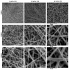
An off-the-shelf vascular graft biomaterial for vascular bypass surgeries is an unmet clinical need. The vascular biomaterial must support cell growth, be non-thrombogenic, minimize intimal hyperplasia, match the structural properties of native vessels, and allow for regeneration of arterial tissue. Electrospun recombinant human tropoelastin (rTE) as a medial component of a vascular graft scaffold was investigated in this study by evaluating its structural properties, as well as its ability to support primary smooth muscle cell adhesion and growth. rTE solutions of 9, 15, and 20 wt% were electrospun into sheets with average fiber diameters of 167 ± 32, 522 ± 67, and 735 ± 270 nm, and average pore sizes of 0.4 ± 0.1, 5.8 ± 4.3, and 4.9 ± 2.4 µm, respectively. Electrospun rTE fibers were cross-linked with disuccinimidyl suberate to produce an insoluble fibrous polymeric recombinant tropoelastin (prTE) biomaterial. Smooth muscle cells attached via integrin binding to the rTE coatings and proliferated on prTE biomaterials at a comparable rate to growth on prTE coated glass, glass alone, and tissue culture plastic. Electrospun tropoelastin demonstrated the cell compatibility and design flexibility required of a graft biomaterial for vascular applications.
Figures


Similar articles
-
Mechanical property characterization of electrospun recombinant human tropoelastin for vascular graft biomaterials.Acta Biomater. 2012 Jan;8(1):225-33. doi: 10.1016/j.actbio.2011.08.001. Epub 2011 Aug 6. Acta Biomater. 2012. PMID: 21846510 Free PMC article.
-
Tropoelastin inhibits intimal hyperplasia of mouse bioresorbable arterial vascular grafts.Acta Biomater. 2017 Apr 1;52:74-80. doi: 10.1016/j.actbio.2016.12.044. Epub 2016 Dec 23. Acta Biomater. 2017. PMID: 28025048 Free PMC article.
-
Alignment of human vascular smooth muscle cells on parallel electrospun synthetic elastin fibers.J Biomed Mater Res A. 2012 Jan;100(1):155-61. doi: 10.1002/jbm.a.33255. Epub 2011 Oct 14. J Biomed Mater Res A. 2012. PMID: 21997972
-
Engineered tropoelastin and elastin-based biomaterials.Adv Protein Chem Struct Biol. 2009;78:1-24. doi: 10.1016/S1876-1623(08)78001-5. Epub 2009 Nov 27. Adv Protein Chem Struct Biol. 2009. PMID: 20663482 Review.
-
Cell-matrix biology in vascular tissue engineering.J Anat. 2006 Oct;209(4):495-502. doi: 10.1111/j.1469-7580.2006.00633.x. J Anat. 2006. PMID: 17005021 Free PMC article. Review.
Cited by
-
Thrombotic responses of endothelial outgrowth cells to protein-coated surfaces.Cells Tissues Organs. 2014;199(4):238-48. doi: 10.1159/000368223. Epub 2015 Jan 16. Cells Tissues Organs. 2014. PMID: 25612682 Free PMC article.
-
Mechanical property characterization of electrospun recombinant human tropoelastin for vascular graft biomaterials.Acta Biomater. 2012 Jan;8(1):225-33. doi: 10.1016/j.actbio.2011.08.001. Epub 2011 Aug 6. Acta Biomater. 2012. PMID: 21846510 Free PMC article.
-
The past, present and future of protein-based materials.Open Biol. 2018 Oct 31;8(10):180113. doi: 10.1098/rsob.180113. Open Biol. 2018. PMID: 30381364 Free PMC article.
-
Tropoelastin enhances nitric oxide production by endothelial cells.Nanomedicine (Lond). 2016 Jun;11(12):1591-7. doi: 10.2217/nnm-2016-0052. Epub 2016 May 13. Nanomedicine (Lond). 2016. PMID: 27175893 Free PMC article.
References
-
- Lloyd-Jones D, Adams RJ, Brown TM, Carnethon M, Dai S, De Simone G, Ferguson TB, Ford E, Furie K, Gillespie C, Go A, Greenlund K, Haase N, Hailpern S, Ho PM, Howard V, Kissela B, Kittner S, Lackland D, Lisabeth L, Marelli A, McDermott MM, Meigs J, Mozaffarian D, Mussolino M, Nichol G, Roger VL, Rosamond W, Sacco R, Sorlie P, Thom T, Wasserthiel-Smoller S, Wong ND, Wylie-Rosett J. Heart disease and stroke statistics--2010 update: a report from the American Heart Association. Circulation. 121(7):e46–e215. - PubMed
-
- Brewster DC. Prosthetic grafts. In: Rutherford RB, editor. Vascular Surgery. 4. Philadelphia: W. B. Saunders; 1995. pp. 492–521.
-
- Abbott WM, Rehring TF. Biologic and synthetic prosthetic materials for vascular conduits. In: Hobson RW, Wilson S, Veith FJ, editors. Vascular Surgery: Principles and Practice. 3. New York: Marcel Dekker; 2004. pp. 611–620.
-
- Sawyer PN, Fitzgerald J, Kaplitt MJ, Sanders RJ, Williams GM, Leather RP, Karmody A, Hallin RW, Taylor R, Fries CC. Ten year experience with the negatively charged glutaraldehyde-tanned vascular graft in peripheral vascular surgery. Initial multicenter trial. Am J Surg. 1987;154(5):533–7. - PubMed
-
- Dardik H, Wengerter K, Qin F, Pangilinan A, Silvestri F, Wolodiger F, Kahn M, Sussman B, Ibrahim IM. Comparative decades of experience with glutaraldehyde-tanned human umbilical cord vein graft for lower limb revascularization: an analysis of 1275 cases. J Vasc Surg. 2002;35(1):64–71. - PubMed
Publication types
MeSH terms
Substances
Grants and funding
LinkOut - more resources
Full Text Sources
Other Literature Sources

