. 2011 May;23(5):1861-75.
doi: 10.1105/tpc.111.085456. Epub 2011 May 17.

A small zinc finger thylakoid protein plays a role in maintenance of photosystem II in Arabidopsis thaliana

Affiliations

A small zinc finger thylakoid protein plays a role in maintenance of photosystem II in Arabidopsis thaliana

Yan Lu et al. Plant Cell. 2011 May.

Abstract

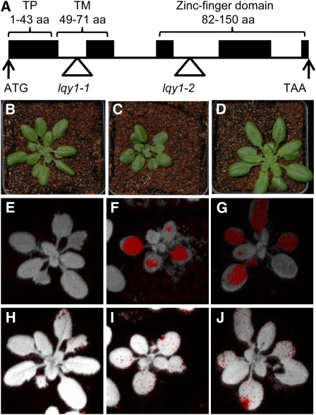
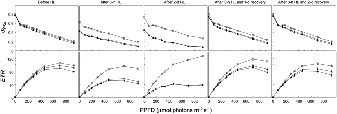
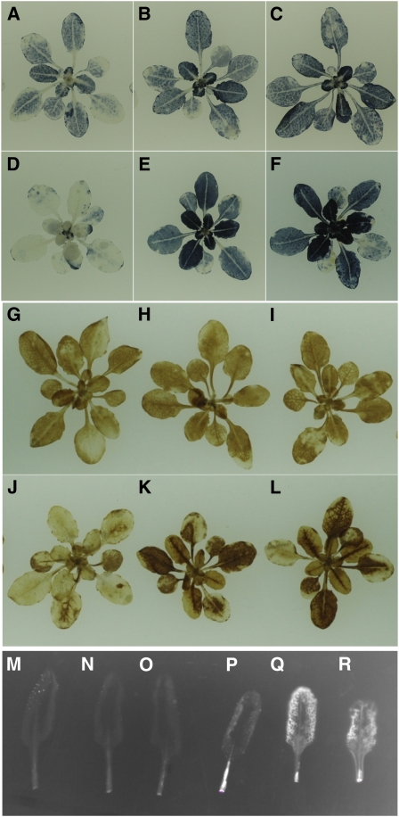
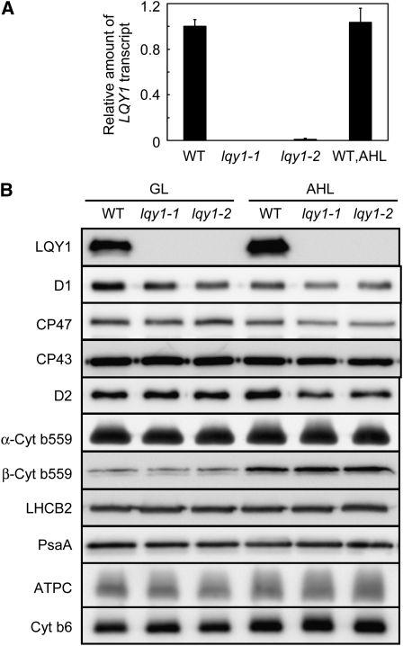
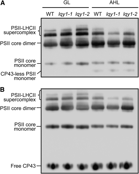
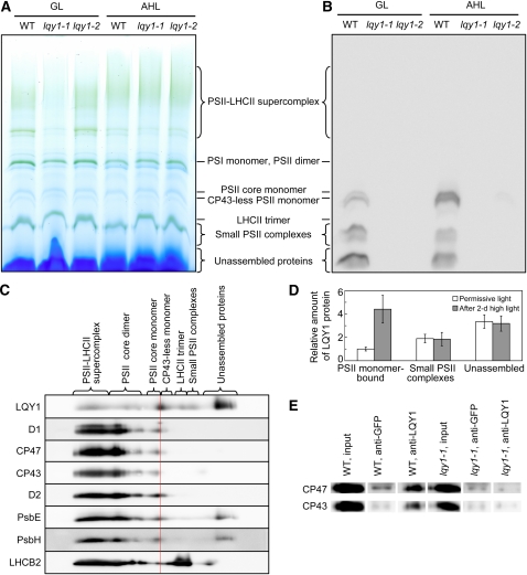
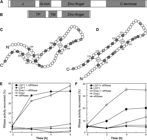
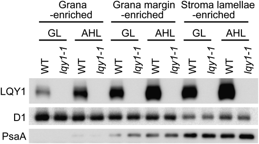
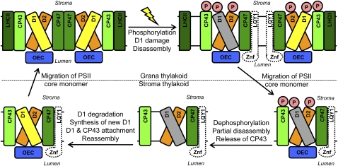
This work identifies LOW QUANTUM YIELD OF PHOTOSYSTEM II1 (LQY1), a Zn finger protein that shows disulfide isomerase activity, interacts with the photosystem II (PSII) core complex, and may act in repair of photodamaged PSII complexes. Two mutants of an unannotated small Zn finger containing a thylakoid membrane protein of Arabidopsis thaliana (At1g75690; LQY1) were found to have a lower quantum yield of PSII photochemistry and reduced PSII electron transport rate following high-light treatment. The mutants dissipate more excess excitation energy via nonphotochemical pathways than wild type, and they also display elevated accumulation of reactive oxygen species under high light. After high-light treatment, the mutants have less PSII-light-harvesting complex II supercomplex than wild-type plants. Analysis of thylakoid membrane protein complexes showed that wild-type LQY1 protein comigrates with the PSII core monomer and the CP43-less PSII monomer (a marker for ongoing PSII repair and reassembly). PSII repair and reassembly involve the breakage and formation of disulfide bonds among PSII proteins. Interestingly, the recombinant LQY1 protein demonstrates a protein disulfide isomerase activity. LQY1 is more abundant in stroma-exposed thylakoids, where key steps of PSII repair and reassembly take place. The absence of the LQY1 protein accelerates turnover and synthesis of PSII reaction center protein D1. These results suggest that the LQY1 protein may be involved in maintaining PSII activity under high light by regulating repair and reassembly of PSII complexes.


Figures

Figure 1.
Figure 1.
Mutations of the LQY1 Gene Cause Altered Chlorophyll Fluorescence Parameters. (A) Gene structure and domains of LQY1 inferred by DNA sequence analysis. Black boxes represent exons and lines represent introns. The start and stop codons are indicated, and the T-DNA insertions in lqy1-1 and lqy1-2 are represented by triangles. aa, amino acids; TM, transmembrane domain; TP, chloroplast transit peptide. (B) to (D) Images of 4-week-old Col wild-type (B), lqy1-1 (C), and lqy1-2 (D) plants under a 12-/12-h photoperiod. (E) to (G) False-color images representing Fv/Fm after a 3-h high-light treatment in 3-week-old Col wild-type (E), lqy1-1 (F), and lqy1-2 (G) plants. Red pixels in (E) to (G) indicate that Fv/Fm is below the cutoff value (0.482). (H) to (J) False-color images representing Fv/Fm after a 3-h high-light treatment and a 2-d recovery period in 3-week-old Col wild-type (H), lqy1-1 (I), and lqy1-2 (J) plants. Red pixels indicate that Fv/Fm is below the cutoff value (0.725).
Figure 2.
Figure 2.
Light–Response Curves of PSII Quantum Yield and ETR in Wild-Type and lqy1 Mutants. The measurements were performed at the following light intensities: 0, 81, 145, 186, 281, 335, 461, 701, and 926 μmol photons m−2 s−1. Data for Col wild-type (open squares), lqy1-1 (filled diamonds), and lqy1-2 (filled triangles) plants are presented as mean ± se (n = 8). HL, high light. Note that lqy1-1 and 1-2 response curves are superimposed in the after 2-d HL ΦPSII and PSII ETR graphs due to their similar phenotypes.
Figure 3.
Figure 3.
Analysis of ROS in Wild-Type and lqy1 Mutants. (A) to (C) NBT staining of O2 in Col wild-type (A), lqy1-1 (B), and lqy1-2 (C) plants under growth light. (D) to (F) NBT staining of O2 in Col wild-type (D), lqy1-1 (E), and lqy1-2 (F) plants after growing under high light for 2 d. (G) to (I) DAB staining of H2O2 in Col wild-type (G), lqy1-1 (H), and lqy1-2 (I) plants under growth light. (J) to (L) DAB staining of H2O2 in Col wild-type (G), lqy1-1 (H), and lqy1-2 (I) plants after growing under high light for 2 d. (M) to (O) Detection of 1O2 with SOSG in Col wild-type (M), lqy1-1 (N), and lqy1-2 (O) plants under growth light. (P) to (R) Detection of 1O2 with SOSG in Col wild-type (P), lqy1-1 (Q), and lqy1-2 (R) plants after growing under high light for 2 d.
Figure 4.
Figure 4.
Analysis of LQY1 Transcript and Thylakoid Membrane Protein Accumulation. (A) Relative amount of the LQY1 transcript determined by quantitative RT-PCR. The amount of LQY1 transcript was normalized by that of the ACT2 transcript (At3g18780). Values (mean ± se, n = 3) are given as the ratio to the amount of LQY1 transcript in wild type (WT) under growth light. For LQY1 transcript levels after a 2-d high-light treatment, only the wild-type value is shown because the transcript was not detectable in either lqy1 mutant. AHL, after 2 d of high light. (B) Representative immunoblots of LQY1 and PSII proteins in wild-type and lqy1 mutants. Thylakoid membrane proteins were separated by SDS-urea-PAGE, electroblotted to a PVDF membrane, and probed with affinity-purified anti-LQY1 or antisera against known thylakoid membrane proteins obtained from Eva-Mari Aro or Agrisera Co. The lanes on each gel were loaded on an equal chlorophyll basis. Cyt, cytochrome; GL, growth light.
Figure 5.
Figure 5.
Immunological Analysis of PSII Complexes Separated by BN-PAGE. (A) A representative immunoblot with anti-D1 antiserum of a BN-PAGE gel. Thylakoid membrane proteins were solubilized with 2% dodecyl β-d-maltoside and separated by BN-PAGE. A sample with an equal amount of chlorophyll (1.5 μg) was loaded in each lane. GL, growth light; AHL, after 2 d of high light; WT, wild type. (B) A representative immunoblot with anti-CP43 antiserum of a BN-PAGE gel as in (A).
Figure 6.
Figure 6.
Evidence for Interaction of LQY1 Protein with PSII Protein Complexes. (A) A representative unstained BN-PAGE gel. Thylakoid membranes were solubilized with 1.5% digitonin and separated on a NativePAGE 3 to 12% Bis-Tris mini gel. A sample with an equal amount of chlorophyll (7 μg) was loaded in each lane. GL, growth light; AHL, after 2 d of high light; WT, wild type. (B) A representative immunoblot with anti-LQY1 antiserum of a gel as in (A). The positions of various PSII protein complexes were determined by immunoblotting with antisera against PSII core proteins D1 and CP43. Little signal is observed in lqy1 mutants, validating the specificity of the antibody. (C) 2-D electrophoresis and immunoblots of PSII protein complexes from Col wild-type leaves after a 2-d high-light treatment. Protein complexes were separated as in (A) by BN-PAGE (first dimension), and proteins in the complexes were separated by SDS-PAGE (second dimension). The 2-D gel was electroblotted and probed with antisera against LQY1 and various PSII proteins. The positions of detected PSII protein complexes are indicated. The red line separates the CP43-less monomer from the PSII core monomer. (D) Relative abundance of LQY1 protein in Col wild-type plants as detected in (B). The values (mean ± se, n = 3) are given as the ratio to PSII monomer-bound LQY1 in Col wild-type plants under growth light. Note that in Figure 6B, the relative intensity of LQY1 in small PSII complexes from wild-type plants decreased after a 2-d high-light treatment. However, this is not seen in Figure 6D, which represents the mean values from three independent BN-PAGE/immunoblots. (E) Immunoprecipitation of CP47 and CP43 by anti-LQY1 antibody. Thylakoid membranes were extracted from Col wild-type and lqy1-1 leaves after a 2-d high-light treatment.
Figure 7.
Figure 7.
The LQY1 Zn Finger Domain Has Protein Disulfide Isomerase Activity. (A) Domains in E. coli DnaJ. The protein contains a J-domain, a Gly-rich motif, a Zn finger domain, and a C-terminal region. (B) Domains inferred from the Arabidopsis LQY1 sequence. The protein contains a transit peptide (TP), a transmembrane region (TM), and a Zn finger domain according to in silico analysis. (C) The topology of the E. coli DnaJ protein (Shi et al., 2005). Cys and Gly residues are shown in gray. Other residues conserved between LQY1 and DnaJ Zn finger are shown in single-letter amino acid codes. (D) Putative topology of Arabidopsis LQY1 protein based on its homology to E. coli DnaJ. (E) Assays for oxidative renaturation of rdRNase by recombinant LQY1 Zn finger. Restoration of activity to inactive reduced and denatured RNase A was measured for LQY1 and positive-control E. coli DnaJ. The values (mean ± se, n = 4) are normalized by the activity of native RNase A. Filled squares, LQY1 and rdRNase; filled diamonds, LQY1, no rdRNase; open squares, DnaJ and rdRNase; open diamonds, DnaJ, no rdRNase; asterisks, rdRNase, no LQY1 or DnaJ. (F) Reductive renaturation of sRNase by recombinant LQY1 Zn finger. The values (mean ± se, n = 4) are normalized as in (E). Filled squares, LQY1 and sRNase; filled diamonds, LQY1, no sRNase; open squares, DnaJ and sRNase; open diamonds, DnaJ, no sRNase; asterisks, sRNase, no LQY1 or DnaJ.
Figure 8.
Figure 8.
Analysis of LQY1 Protein in Grana-, Grana Margin-, and Stroma Lamellae-Enriched Thylakoid Membranes. Thylakoid membranes were solubilized by 0.4% digitonin and fractionated by ultracentrifugation. Fractionated thylakoid membrane proteins were separated by SDS-Urea-PAGE and analyzed by immunoblots. The lanes on each gel were loaded on an equal chlorophyll basis. GL, growth light; AHL, after 2-d high light; WT, wild type.
Figure 9.
Figure 9.
PSII Repair and Reassembly and Hypothetical Role of LQY1. This schematic diagram for high-light induced PSII repair and reassembly is modified from Mulo et al. (2008). Undamaged D1 protein is yellow-filled and damaged D1 protein is gray-filled. The thylakoid membrane-localized LQY1 protein, schematically drawn as an unfilled and dashed shape, comigrates with PSII core monomer and CP43-less core monomer on BN-PAGE gels. The Zn finger domain (Znf) in LQY1 is predicted to locate at the lumen side of thylakoid membrane. The presence of LQY1 in these complexes, its mutant phenotype, and protein disulfide isomerase activity are consistent with a role in PSII photodamage repair. The specific proteins with which it interacts and the substrates for its disulfide isomerase activity remain to be determined. OEC, oxygen evolving complex.

References

    1. Ajjawi I., Lu Y., Savage L.J., Bell S.M., Last R.L. (2010). Large-scale reverse genetics in Arabidopsis: Case studies from the Chloroplast 2010 Project. Plant Physiol. 152: 529–540 - PMC - PubMed
    1. Alonso J.M., et al. (2003). Genome-wide insertional mutagenesis of Arabidopsis thaliana. Science 301: 653–657 - PubMed
    1. Aro E.-M., Suorsa M., Rokka A., Allahverdiyeva Y., Paakkarinen V., Saleem A., Battchikova N., Rintamäki E. (2005). Dynamics of photosystem II: A proteomic approach to thylakoid protein complexes. J. Exp. Bot. 56: 347–356 - PubMed
    1. Baena-González E., Aro E.M. (2002). Biogenesis, assembly and turnover of photosystem II units. Philos. Trans. R. Soc. Lond. B Biol. Sci. 357: 1451–1459, discussion 1459–1460 - PMC - PubMed
    1. Baker N.R., Harbinson J., Kramer D.M. (2007). Determining the limitations and regulation of photosynthetic energy transduction in leaves. Plant Cell Environ. 30: 1107–1125 - PubMed

Publication types

MeSH terms