. 2011:2:315.
doi: 10.1038/ncomms1308.

The neural circuits and sensory channels mediating harsh touch sensation in Caenorhabditis elegans

Affiliations

The neural circuits and sensory channels mediating harsh touch sensation in Caenorhabditis elegans

Wei Li et al. Nat Commun. 2011.

Abstract

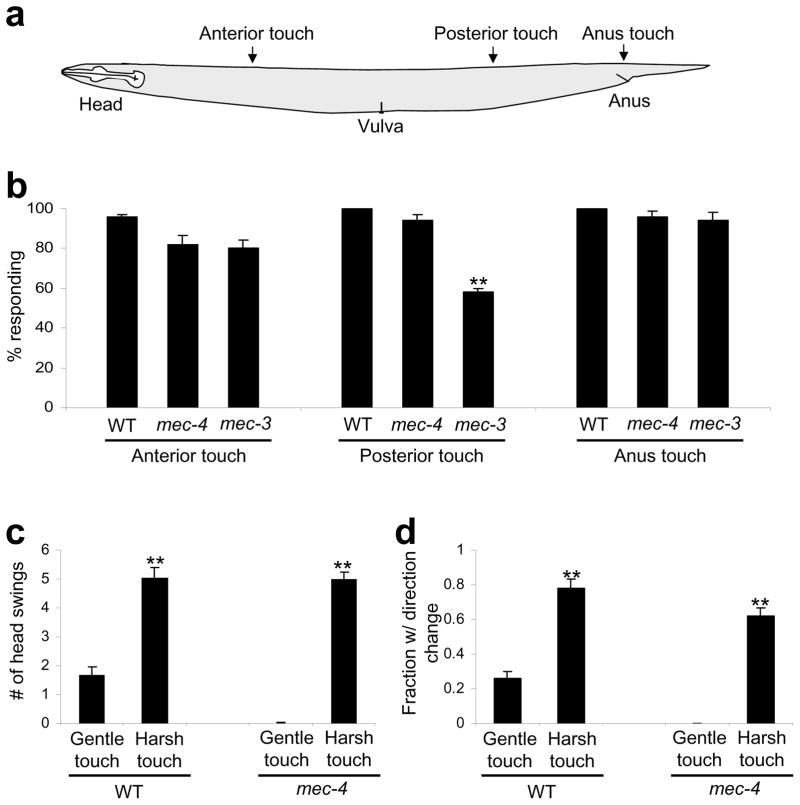
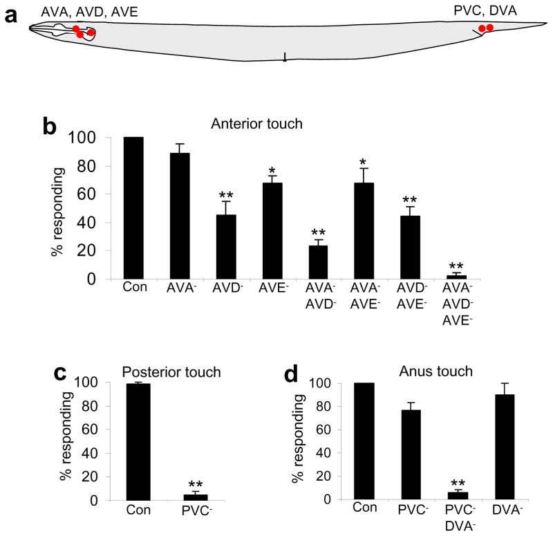
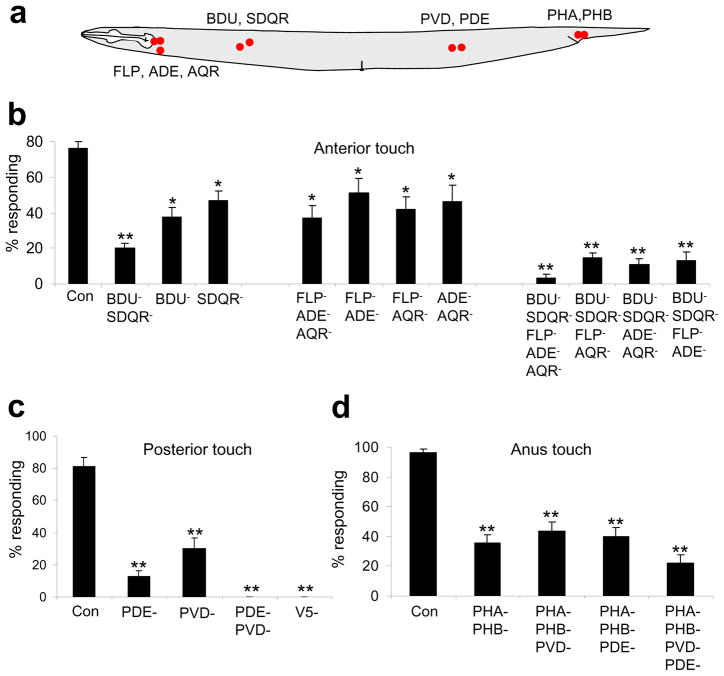
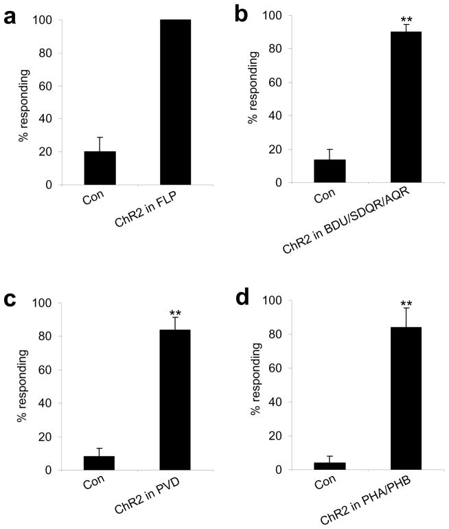
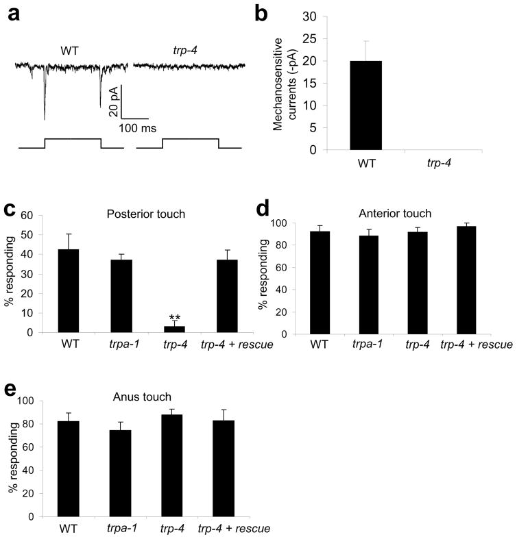
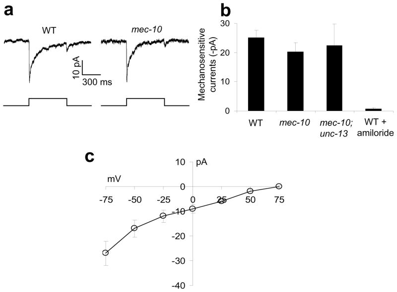
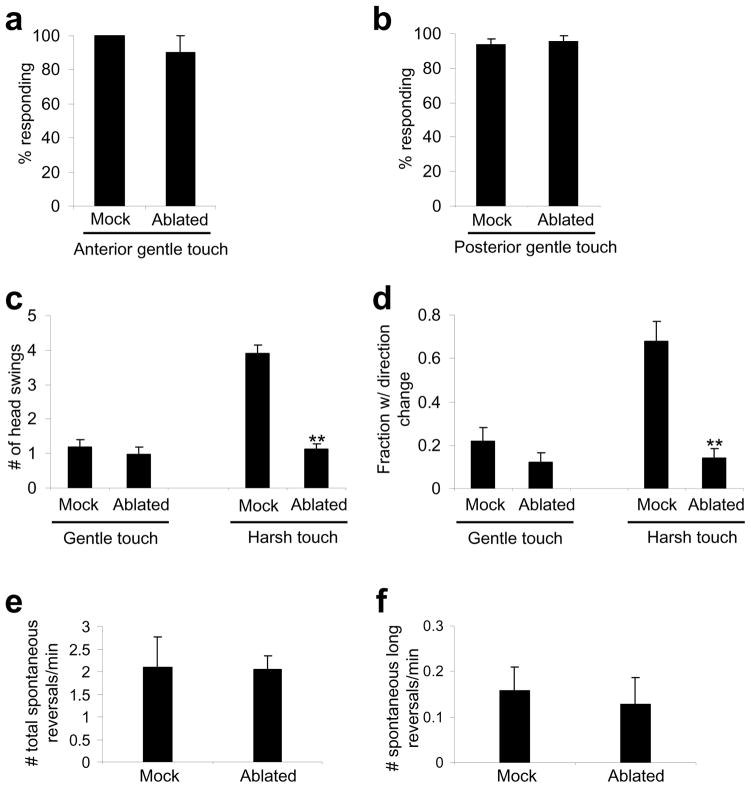
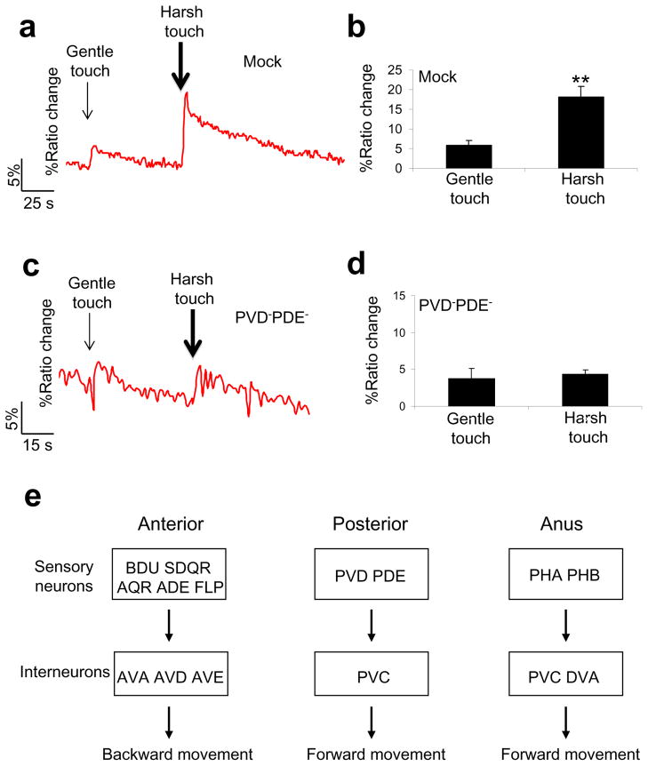
Most animals can distinguish two distinct types of touch stimuli: gentle (innocuous) and harsh (noxious/painful) touch, however, the underlying mechanisms are not well understood. Caenorhabditis elegans is a useful model for the study of gentle touch sensation. However, little is known about harsh touch sensation in this organism. Here we characterize harsh touch sensation in C. elegans. We show that C. elegans exhibits differential behavioural responses to harsh touch and gentle touch. Laser ablations identify distinct sets of sensory neurons and interneurons required for harsh touch sensation at different body segments. Optogenetic stimulation of the circuitry can drive behaviour. Patch-clamp recordings reveal that TRP family and amiloride-sensitive Na(+) channels mediate touch-evoked currents in different sensory neurons. Our work identifies the neural circuits and characterizes the sensory channels mediating harsh touch sensation in C. elegans, establishing it as a genetic model for studying this sensory modality.


Conflict of interest statement

Competing financial interests: The authors declare no competing financial interests.

Figures

Figure 1
Figure 1. Animals exhibit differential behavioral responses to harsh touch and gentle touch
(a) A schematic illustrating the location of harsh touch stimuli delivered to different body segments. (b) Response of WT, mec-4(e1611) and mec-3(e1338) worms to anterior, posterior and anus harsh touch. mec-4(e1611) and mec-3(e1338) worms were outcrossed for six and four times, respectively. The non-outcrossed CGC strain CB1338 of mec-3(e1338) was a bit unhealthy and showed reduced responses (Supplementary Figure S1). n=10. **p<0.0001 (ANOVA with Bonferroni test). Error bars: SEM. (c) Animals respond to harsh touch more robustly than gentle touch. The number of head swings of backward movement triggered by anterior gentle and harsh touch was scored. **p<0.0001 (t test; harsh touch vs. gentle touch). However, harsh touch data from mec-4(e1611) mutant and wild-type under were not significantly different. n=10. Error bars: SEM. (d) The frequency of direction change following backward movement triggered by anterior harsh touch and gentle touch. A direction change was defined as a ≥90 degree turn from the previous locomotion direction. **p<0.0001 (t test; harsh touch vs. gentle touch). However, harsh touch data from mec-4(e1611) mutant strain and wild-type were not significantly different. n=10. Error bars: SEM.
Figure 2
Figure 2. Identification of the interneurons required for harsh touch behavior
(a) A schematic illustrating the position of interneuron cell bodies. (b) AVA, AVD and AVE are required for mediating anterior but not posterior or anus harsh touch response. *p<0.02. **p<0.0001 (ANOVA with Bonferroni test compared to control). n≥5. Error bars: SEM. (c) PVC is required for mediating posterior but not anterior or anus harsh touch response. **p<0.0001 (t test). n≥12. Error bars: SEM. (d) PVC and DVA together are required for mediating anus harsh touch response. **p<0.0001 (ANOVA with Bonferroni test compared to control). n≥4. Error bars: SEM.
Figure 3
Figure 3. Identification of the sensory neurons required for harsh touch behavior
(a) A schematic illustrating the location of sensory neuron cell bodies. (b) Two groups of sensory neurons are required for mediating anterior harsh touch response. The anterior gentle touch sensory neurons ALM and AVM were killed in all ablations. *p<0.03. **p<0.0001 (ANOVA with Bonferroni test compared to control). n≥9. Error bars: SEM. (c) PVD and PDE together are required for mediating posterior harsh touch response. The posterior gentle touch sensory neuron PLM was killed in all ablations. V5 is the precursor cell of PVD and PDE. **p<0.0001 (ANOVA with Bonferroni test compared to control). n≥7. Error bars: SEM. (d) PHA/PHB are important for mediating anus harsh touch response. PLM was killed in all ablations. **p<0.0001 (ANOVA with Bonferroni test compared to control). n≥9. Error bars: SEM.
Figure 4
Figure 4. Optogenetic stimulation of sensory neurons by ChR2 can drive behavioral responses
(a) ChR2 stimulation of FLP can drive backward movement. ChR2 was expressed in FLP as a transgene under the mec-3 promoter in the lite-1(xu7)mec-4(e1611) background. The mec-3 promoter labels FLP, PVD and gentle touch neurons. lite-1(xu7) abolishes intrinsic phototaxis response. mec-4(e1611) eliminates all gentle touch neurons. We thus analyzed FLP-positive animals in which PVD was killed by laser. Control animals were transgene-free siblings. **p<0.0001 (t test). n≥5. Error bars: SEM. (b) ChR2 stimulation of BDU, SDQR and AQR can drive backward movement. ChR2 was expressed as a transgene under the gcy-35 promoter in the lite-1(xu7) background. This promoter also labels URX and AVM in the head and anterior body , so we analyzed animals with URX and AVM ablated. Control animals were transgene-free siblings. **p<0.0001 (t test). n≥6. Error bars: SEM. (c) ChR2 stimulation of PVD can drive forward movement. ChR2 was expressed in PVD as a transgene under the mec-3 promoter in the lite-1(xu7)mec-4(e1611) background. We analyzed PVD-positive animals with FLP killed by laser. Control animals were transgene-free siblings. **p<0.0001 (t test). n≥5. Error bars: SEM. (d) ChR2 stimulation of PHA/PHB can drive forward movement. ChR2 was expressed as a transgene under the ocr-2 promoter in the lite-1(xu7) background . To avoid turning on the head neurons labeled by the ocr-2 promoter, we directed light pulses to the animal tail. Control animals were transgene-free siblings. **p<0.0001 (t test). n≥5. Error bars: SEM.
Figure 5
Figure 5. The TRPN channel TRP-4 regulates posterior harsh touch response
(a) Touch evokes mechanosensitive currents in PDE in a TRP-4-dependent manner. Clamping voltage: −75 mV. Touch was directed to the cilium of PDE. Displacement: 10 μm. 1 μm displacement was sufficient to evoke mechanosensitive currents in PDE. (b) Bar graph summarizing the data in (a). n≥5. Error bars: SEM. (c) TRP-4 is required for posterior harsh touch behavioral response mediated by PDE. To rescue the defect, wild-type trp-4 cDNA was expressed as a transgene in PDE under the dat-1 promoter in trp-4(sy695) mutant animals, . PVD and PLM were both killed in each genotype. **p<0.0001 (ANOVA with Bonferroni test compared to control). n≥7. Error bars: SEM. (d) Responses to anterior harsh touch in worms described in (c). (e) Responses to anus harsh touch in worms described in (c).
Figure 6
Figure 6. An amiloride-sensitive channel(s) mediates mechanosensitive currents in PVD
(a) Mechanical stimulation evokes mechanosensitive currents in PVD. Such currents persisted in mec-10(tm1552). Clamping voltage: −75 mV. Touch was directed to the primary dendrite of PVD. Displacement: 20 μm. A 10 μm displacement was needed to evoke mechanosensitive currents in PVD, a much higher threshold than that in PDE. PVD in mec-10(tm1552) animals displayed a similar sensitivity (threshold). (b) Bar graphs. Touch-evoked mechanosensitive currents in PVD were sensitive to amiloride (200 μM) but persisted in mec-10(tm1552), as well as in mec-10(tm1552); unc-13(e51) double mutant animals. n≥5. Error bars: SEM. (c) I–V relations of mechanosensitive currents in PVD. n≥5. Error bars: SEM.
Figure 7
Figure 7. Animals lacking harsh touch sensory neurons can no longer distinguish harsh touch from gentle touch
(a) Worms lacking anterior harsh touch sensory neurons retain sensitivity to anterior gentle touch. All five anterior sensory neurons (BDU, SDQR, FLP, ADE and AQR) were ablated. Anterior gentle touch neurons (ALM and AVM) were not killed. n=10. Error bars: SEM. (b) Worms lacking posterior harsh touch sensory neurons retain sensitivity to posterior gentle touch. PDE and PVD were ablated. The posterior gentle touch neuron PLM was not killed. n=10. Error bars: SEM. (c) Worms lacking harsh touch sensory neurons responded similarly to harsh touch and gentle touch. All anterior harsh touch sensory neurons were ablated, and the ablated animals were assayed for anterior gentle and harsh touch responses. The number of head swings of backward movement triggered by anterior harsh and gentle touch was scored. Gentle touch sensory neurons were not killed. n=10. **p<0.0001 (ANOVA with Bonferroni test compared to mock). Error bars: SEM. (d) The same worms in (c) were scored for the frequency of direction change following backward movement. n=10. **p<0.0001 (ANOVA with Bonferroni test compared to mock). Error bars: SEM. (e) Animals lacking all anterior harsh touch sensory neurons show a normal frequency of spontaneous reversals. n=10. Error bars: SEM. (f) Animals lacking all anterior harsh touch sensory neurons show a normal frequency of long reversals during spontaneous locomotion. Long reversals are defined as backward movement events with >3 head swings. n=10. Error bars: SEM.
Figure 8
Figure 8. Differential stimulation of the interneuron PVC by gentle and harsh touch
(a) Harsh touch evokes a greater calcium response in PVC. Gentle touch (5μm at 20 Hz; 3 s) and harsh touch (20 μm at 20 Hz; 3 s) were sequentially delivered to the posterior body of immobilized animals. (b) Bar graph summarizing the data in (a). **p<0.0001 (t test). n=21. Error bars: SEM. (c) Animals lacking harsh touch sensory neurons responded similarly to harsh and gentle touch in PVC. (d) Bar graph summarizing the data in (c). n=5. Error bars: SEM. (e) A diagram summarizing the neural circuits underlying harsh touch sensation. The sensory neurons and interneurons required for harsh touch sensation in each body segment are listed.

References

    1. Bounoutas A, Chalfie M. Touch sensitivity in Caenorhabditis elegans. Pflugers Arch. 2007;454:691–702. - PubMed
    1. Purves D, et al. Sensation and sensory processsing. In: Fitzpatrick D, editor. Neuroscience. Sinauer Associates, Inc; Sunderland: 2008. pp. 207–393.
    1. Bargmann CI. Chemosensation in C. elegans. WormBook; 2006. pp. 1–29. - PMC - PubMed
    1. Ward A, Liu J, Feng Z, Xu XZ. Light-sensitive neurons and channels mediate phototaxis in C. elegans. Nature Neurosci. 2008;11:916–922. - PMC - PubMed
    1. Liu J, et al. C. elegans phototransduction requires a G protein–dependent cGMP pathway and a taste receptor homolog. Nature neuroscience. 2010;13:715–722. - PMC - PubMed

Publication types

MeSH terms

Substances