. 2011:2:318.
doi: 10.1038/ncomms1317.

IKKβ regulates essential functions of the vascular endothelium through kinase-dependent and -independent pathways

Affiliations
Free PMC article

IKKβ regulates essential functions of the vascular endothelium through kinase-dependent and -independent pathways

Noboru Ashida et al. Nat Commun. 2011.
Free PMC article

Abstract

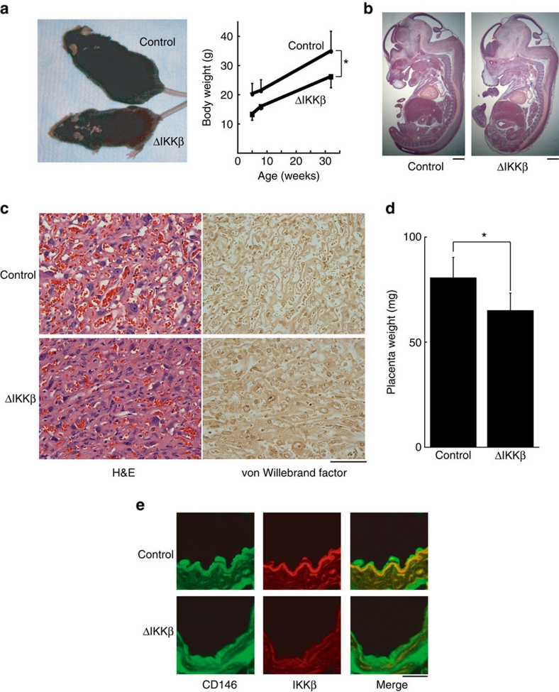
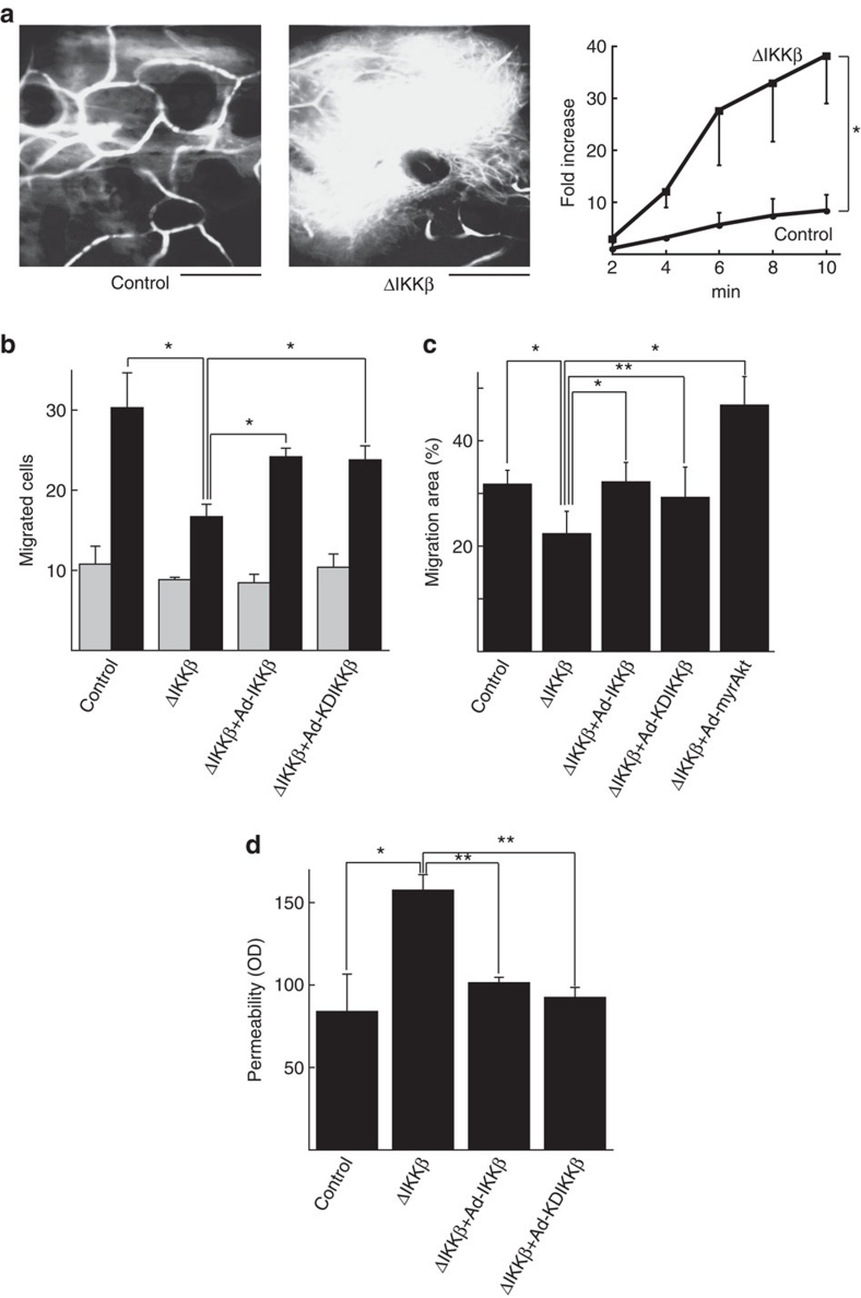
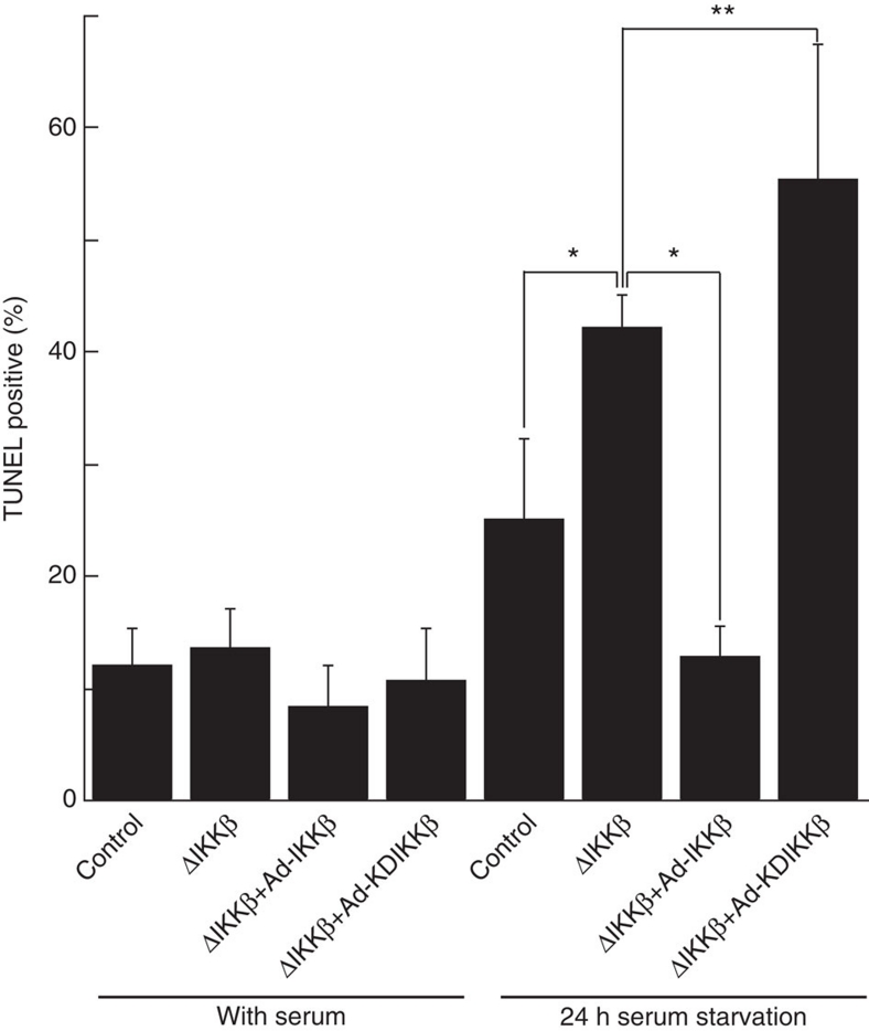
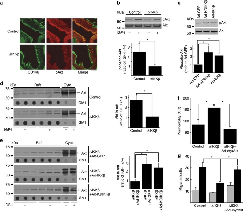
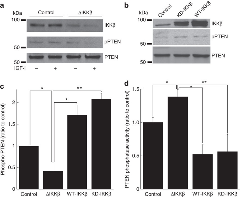
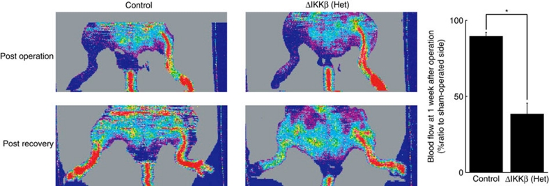
Vascular endothelium provides a selective barrier between the blood and tissues, participates in wound healing and angiogenesis, and regulates tissue recruitment of inflammatory cells. Nuclear factor (NF)-κB transcription factors are pivotal regulators of survival and inflammation, and have been suggested as potential therapeutic targets in cancer and inflammatory diseases. Here we show that mice lacking IKKβ, the primary kinase mediating NF-κB activation, are smaller than littermates and born at less than the expected Mendelian frequency in association with hypotrophic and hypovascular placentae. IKKβ-deleted endothelium manifests increased vascular permeability and reduced migration. Surprisingly, we find that these defects result from loss of kinase-independent effects of IKKβ on activation of the serine-threonine kinase, Akt. Together, these data demonstrate essential roles for IKKβ in regulating endothelial permeability and migration, as well as an unanticipated connection between IKKβ and Akt signalling.


Figures

Figure 1
Figure 1. Phenotype of endothelial-specific IKKβ knockout mice.
(a) Photograph of littermate wild type ('control') and IKKβΔEC ('ΔIKKβ') mice and body weight±s.d. shown from mice of each genotype at the indicated age (n=3/each group, *P<0.005 in repeated two-way ANOVA). (b) H&E staining of embryos at E14. Scale bars: 1 mm. (c) Staining of placentae by H&E and immunohistochemistry for von Willebrand Factor at E14. Scale bars: 10 μm. (d) Placenta weight from mice of each genotype (n=6 in IKKβΔEC and n=3 in control mice, *P<0.05 in two-sided Student's t-test. Error bars:±s.d.). (e) Confocal microscopy images of vascular endothelium in IKKβΔEC and control mice immunostained with antibody for CD146 and IKKβ. Scale bars: 100 μm.
Figure 2
Figure 2. Permeability and migration in IKKβ-manipulated endothelial cells in vivo and in vitro.
(a) In vivo microscopy images of ear microvessels after injection of Evans blue dye. Representative image at 10 min (top) and cumulative data±s.e. from three independent experiments (bottom) are shown (*P<0.005 in repeated two-way ANOVA). Scale bars: 100 μm. See also Supplementary Movie 1 online. (b) In vitro migration to VEGF of endothelial cells in which IKKβ is deleted or WT/-IKKβ is expressed. Cumulative data±s.e. from four independent experiments are shown. Grey bars: VEGF (−), black bars: VEGF (+). *P<0.05 in two-sided Student's t-test. (c) Wound-healing assay. Migration area was accumulated from six separated observation fields. Representative experiment is shown from three independent experiments total. *P<0.005 in two-sided Student's t-test. **P<0.05 in two-sided Student's t-test. Error bars: ±s.d. (d) In vitro permeability of endothelial cells in which IKKβ is deleted or WT/KD-IKKβ is expressed. Cumulative data±s.e. from three independent experiments are shown. *P<0.05 in two-sided Student's t-test. **P<0.005 in two-sided Student's t-test. OD, optical density.
Figure 3
Figure 3. Effect of IKKβ on apoptosis induced by 24 h serum starvation.
Primary cultured endothelial cells from lungs of IKKβflox/flox mice with infection of Ad-GFP, Cre, IKKβ or KDIKKβ were serum-starved for 24 h. TUNEL positive cells were counted at seven independent fields (mean cell number: 241.5/field). *P<0.0005 in two-sided Student's t-test, **P<0.05 in two-sided Student's t-test. Error bars: ±s.d.
Figure 4
Figure 4. Akt phosphorylation and localization in IKKβ-manipulated endothelial cells in vivo and in vitro.
(a) Confocal microscopy images of endothelial cells in IKKβΔEC mice immunostained with antibody for CD146 and pAkt. Scale bar: 20 μm. (b) Representative immunoblots for pAkt and Akt in IKKβ-deleted endothelial cells treated with/without IGF-I and its cumulative quantitative densitometry data±s.d. from three independent experiments (*P<0.05 in two-sided Student's t-test). (c) Representative immunoblots for pAkt and Akt in endothelial cells overexpressed with WT- or KD-IKKβ and its cumulative quantitative densitometry data±s.d. from three independent experiments (*P<0.05 in two-sided Student's t-test). (d) Representative Akt immunoblots of fractions from sucrose gradient centrifugation of IKKβ-deleted endothelial cells treated with/without IGF-I and its cumulative quantitative densitometry data±s.e. from three independent experiments (*P<0.05 in two-sided Student's t-test). Ganglioside GM1 is marker for lipid raft fractions. (e) Representative Akt immunoblots of fractions from sucrose gradient centrifugation of IKKβ-deleted endothelial cells expressed with either WT- or KD-IKKβ treated with/without IGF-I and its cumulative quantitative densitometry data±s.e. (n=5 for ΔIKKβ+Ad-GFP; n=3 for ΔIKKβ+Ad-IKKβ; n=5 for ΔIKKβ+Ad-KDIKKβ; *P<0.05 in two-sided Student's t-test). Ganglioside GM1 is marker for lipid raft fractions. (f, g) In vitro permeability (f) and migration to VEGF (g) in endothelial cells in which IKKβ is deleted or myr-Akt is expressed. Gray bars: VEGF (−), black bars: VEGF (+) for (g). Each panel is cumulative data±s.e. from three independent experiments (*P<0.05 in two-sided Student's t-test).
Figure 5
Figure 5. PTEN phosphorylation and phosphatase activity in IKKβ-manipulated endothelial cells.
(ac) Representative immunoblots for IKKβ, pPTEN or PTEN in IKKβ-deleted endothelial cells (a) or WT/KD IKKβ expressed endothelial cells (b) and its cumulative quantitative densitometry data±s.d. from three independent experiments (c) (*P<0.05 in two-sided Student's t-test, **P<0.0005 in two-sided Student's t-test). (d) Phosphatase activity of PTEN in IKKβ-deleted or WT/KD IKKβ expressed endothelial cells. Cumulative data±s.d. from four independent experiments are shown (*P<0.005 in two-sided Student's t-test, **P<0.05 in two-sided Student's t-test).
Figure 6
Figure 6. Ischemia-induced neovascularization in vivo.
Representative images of blood flow after ligation of common femoral artery, and quantified cumulative data±s.d. from six animals (3 Cre-negative, 3 heterozygous IKKβΔEC mice. *P<0.0005 in two-sided Student's t-test).

References

    1. Karin M. Nuclear factor-kappaB in cancer development and progression. Nature 441, 431–436 (2006). - PubMed
    1. Zandi E., Rothwarf D. M., Delhase M., Hayakawa M. & Karin M. The IkappaB kinase complex (IKK) contains two kinase subunits, IKKalpha and IKKbeta, necessary for IkappaB phosphorylation and NF-kappaB activation. Cell 91, 243–252 (1997). - PubMed
    1. Mercurio F. et al.. IKK-1 and IKK-2: cytokine-activated IkappaB kinases essential for NF- kappaB activation. Science 278, 860–866 (1997). - PubMed
    1. Rothwarf D. M., Zandi E., Natoli G. & Karin M. IKK-gamma is an essential regulatory subunit of the IkappaB kinase complex. Nature 395, 297–300 (1998). - PubMed
    1. Hu Y. et al.. IKKalpha controls formation of the epidermis independently of NF-kappaB. Nature 410, 710–714 (2001). - PubMed

Publication types

Substances

LinkOut - more resources