Review
. 2011 Jun;12(6):359-66.
doi: 10.1038/nrn3039. Epub 2011 May 18.

Ageing as a primary risk factor for Parkinson's disease: evidence from studies of non-human primates

Affiliations
Review

Ageing as a primary risk factor for Parkinson's disease: evidence from studies of non-human primates

Timothy J Collier et al. Nat Rev Neurosci. 2011 Jun.

Abstract

Ageing is the greatest risk factor for the development of Parkinson's disease. However, the current dogma holds that cellular mechanisms that are associated with ageing of midbrain dopamine neurons and those that are related to dopamine neuron degeneration in Parkinson's disease are unrelated. We propose, based on evidence from studies of non-human primates, that normal ageing and the degeneration of dopamine neurons in Parkinson's disease are linked by the same cellular mechanisms and, therefore, that markers of cellular risk factors accumulate with age in a pattern that mimics the pattern of degeneration observed in Parkinson's disease. We contend that ageing induces a pre-parkinsonian state, and that the cellular mechanisms of dopamine neuron demise during normal ageing are accelerated or exaggerated in Parkinson's disease through a combination of genetic and environmental factors.


Conflict of interest statement

Competing interests statement

J.H.K. declares competing financial interests: see web version for details. The remaining authors declare no competing financial interests.

Figures

Figure 1
Figure 1. Regional differences in vulnerability to degeneration in Parkinson’s disease and models of parkinsonism
Dopamine neurons of the ventral midbrain vary in their susceptibility to degeneration, with neurons of the ventral tier of substantia nigra (vtSN, shown in red) being the most vulnerable, the dorsal tier of substantia nigra (dtSN, shown in yellow) being less vulnerable, and the adjacent cells of the ventral tegmental area (VTA, shown in green) being the least vulnerable. The low magnification image is a coronal hemisection through the ventral midbrain of a rhesus monkey stained with cresyl violet. The expanded view is stained for tyrosine hydroxylase as a marker for dopamine neurons. The red, yellow and green areas correspond to the areas compared for cell counts and expression of immunocytochemical markers in the studies that are reviewed in this article.
Figure 2
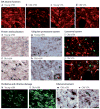
Figure 2. The pattern of ageing-related changes in markers of cellular mechanisms
With advancing age, dopamine (DA) neurons in the ventral tier of the substantia nigra (vtSN) — the population that is most vulnerable to degeneration in Parkinson’s disease — show changes with ageing. a,b,c,d | Age-related decline in tyrosine hydroxylase staining (shown in red) in vtSN neurons but not in vental tegmental area (VTA) neurons. e,f | Accumulation of cytoplasmic α-synuclein (shown in brown). Tyrosine hydroxylase staining is shown in grey. Arrows show examples of cytoplasmic α-synuclein in aged vtSN. g,h | Increased numbers of Marinesco bodies, characterized by cytoplasmic inclusions of ubiquitin (shown in red). Tyrosine hydroxylase staining is shown in grey. The inset (in part h) is a higher magnification view of a tyrosine hydroxylase immunoreactive neuron of the vtSN exhibiting multiple Marinesco bodies. i,j | No accumulation of lipofuscin (shown in green). Tyrosine hydroxylase staining is shown in red, colocalization of lipofuscin and tyrosine hydroxylase is shown in yellow. Note that virtually all lipofuscin staining in the vtSN is not in dopamine neurons, whereas colocalization is apparent in aged VTA neurons. k,l | Accumulation of 3-nitrotyrosine (shown in green). m,n | Greater microglial reactivity in aged vtSN neurons than in aged VTA neurons, shown by greater staining for HLA class II histocompatibility antigen, DR α-chain (HLA-DRA; a marker for microglia), shown in brown. UPS, ubiquitin–proteasome system. Parts e and f are reproduced, with permission, from REF. © (2007) Elsevier. Parts k and l are reproduced, with permission, from REF. © (2008) Blackwell Publishing.
Figure 3
Figure 3. The stochastic acceleration hypothesis
A revised hypothesis of the relationship between ageing and Parkinson’s disease (PD) as they affect the biology of midbrain dopamine (DA) neurons. The hypothesis incorporates evidence that supports the involvement of common cellular mechanisms involved in dopamine neuron dysfunction in ageing, and degeneration in Parkinson’s disease. a | The effects of these altered cellular mechanisms as they accumulate during normal ageing result in parkinsonian dopamine neuron dysfunction, either very late in life or not at all (shown by the light grey line). However, when these same cellular mechanisms are accelerated by specific, individually determined factors, parkinsonism emerges earlier in the lifespan (shown by the dark grey line). b | The hypothesis contends that the cellular mechanisms that threaten dopamine neuron function are identical, but not linked in an orderly cascade of cause and effect, and instead can contribute to varying degrees and combine in patient-specific patterns, thus fulfilling the definition of a stochastic interaction: incorporating elements of randomness with directionality towards dopamine neuron dysfunction. Light grey double-ended arrows show cellular events in normal ageing. Thicker, dark grey double-ended arrows show accelerated cellular events in PD. UPS, ubiquitin–proteasome system.

References

    1. Bennett DA, et al. Prevalence of parkinsonian signs and associated mortality in a community population of older people. N Engl J Med. 1996;334:71–76. - PubMed
    1. Morens DM, et al. Epidemiologic observations on Parkinson’s disease: incidence and mortality in a prospective study of middle aged men. Neurology. 1996;46:1044–1050. - PubMed
    1. Tanner CM, Goldman SM. Epidemiology of Parkinson’s disease. Neurol Clin. 1996;14:317–335. - PMC - PubMed
    1. Fearnley JM, Lees AJ. Ageing and Parkinson’s disease: substantia nigra regional selectivity. Brain. 1991;114:2283–2301. - PubMed
    1. Gibb WR, Lees AJ. Anatomy, pigmentation, ventral and dorsal subpopulations of the substantia nigra, and differential cell death in Parkinson’s disease. J Neurol Neurosurg Psychiatr. 1991;54:388–396. - PMC - PubMed