. 2009 Dec 7;2(4):9-23.
doi: 10.3822/ijtmb.v2i4.62.

The architecture of the connective tissue in the musculoskeletal system-an often overlooked functional parameter as to proprioception in the locomotor apparatus

Affiliations

The architecture of the connective tissue in the musculoskeletal system-an often overlooked functional parameter as to proprioception in the locomotor apparatus

Jaap van der Wal. Int J Ther Massage Bodywork. .

Abstract

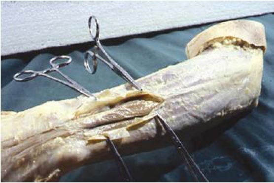
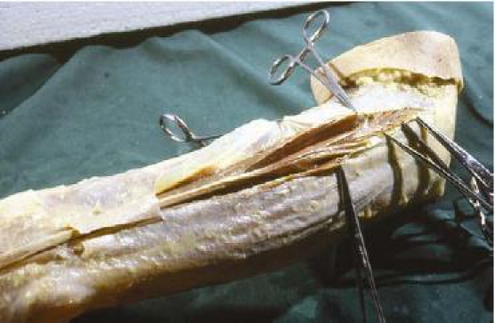
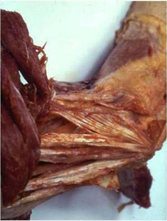
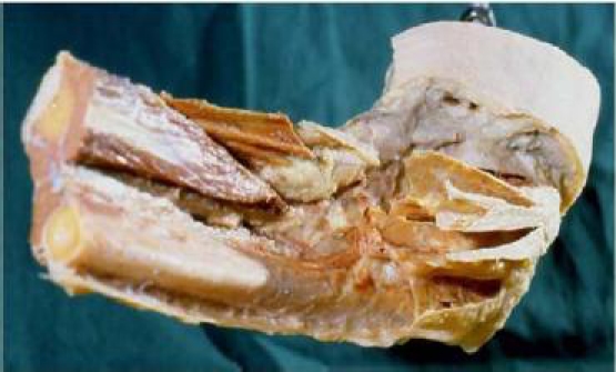
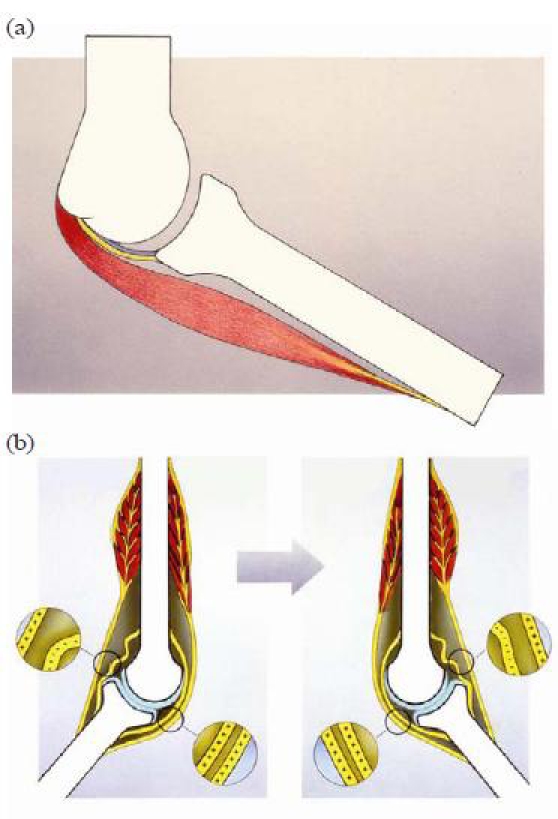
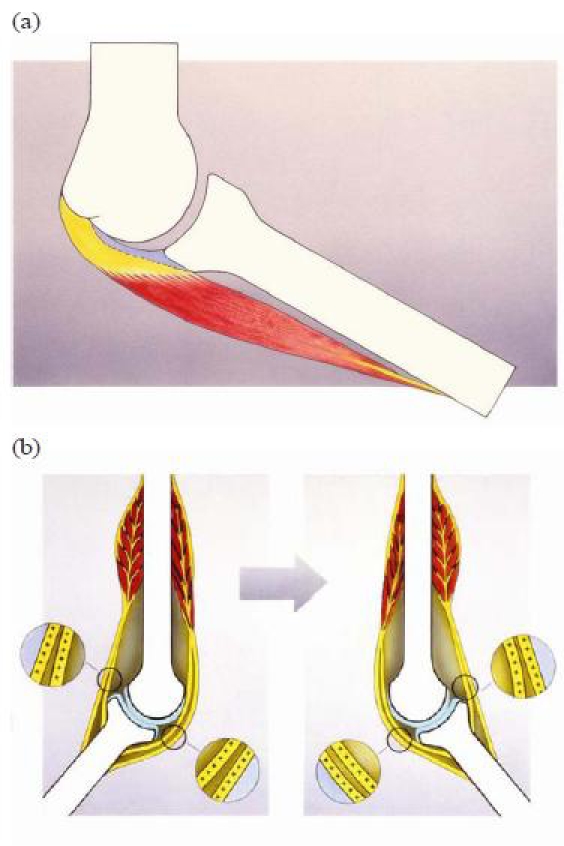
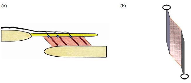
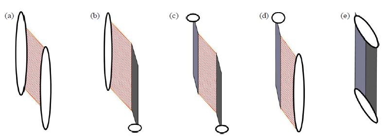
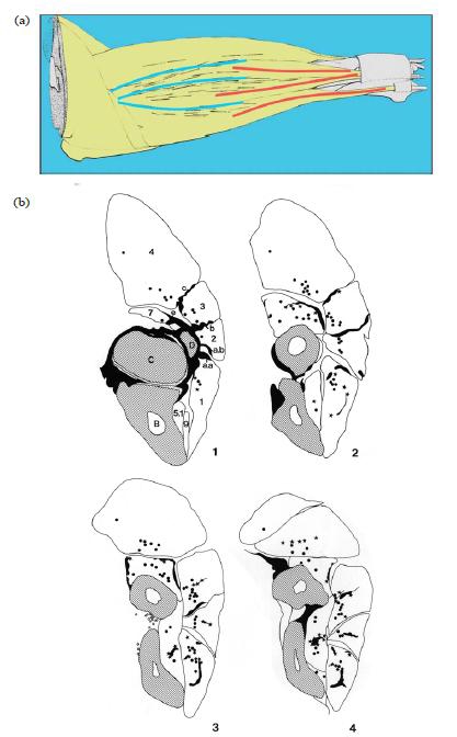
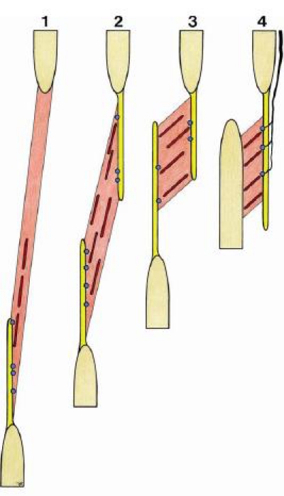
The architecture of the connective tissue, including structures such as fasciae, sheaths, and membranes, is more important for understanding functional meaning than is more traditional anatomy, whose anatomical dissection method neglects and denies the continuity of the connective tissue as integrating matrix of the body.The connective tissue anatomy and architecture exhibits two functional tendencies that are present in all areas of the body in different ways and relationships. In body cavities, the "disconnecting" quality of shaping space enables mobility; between organs and body parts, the "connecting" dimension enables functional mechanical interactions. In the musculoskeletal system, those two features of the connective tissue are also present. They cannot be found by the usual analytic dissection procedures. An architectural description is necessary.This article uses such a methodologic approach and gives such a description for the lateral elbow region. The result is an alternative architectural view of the anatomic substrate involved in the transmission and conveyance of forces over synovial joints. An architectural description of the muscular and connective tissue organized in series with each other to enable the transmission of forces over these dynamic entities is more appropriate than is the classical concept of "passive" force-guiding structures such as ligaments organized in parallel to actively force-transmitting structures such as muscles with tendons.The discrimination between so-called joint receptors and muscle receptors is an artificial distinction when function is considered. Mechanoreceptors, also the so-called muscle receptors, are arranged in the context of force circumstances-that is, of the architecture of muscle and connective tissue rather than of the classical anatomic structures such as muscle, capsules, and ligaments. In the lateral cubital region of the rat, a spectrum of mechanosensitive substrate occurs at the transitional areas between regular dense connective tissue layers and the muscle fascicles organized in series with them. This substrate exhibits features of type and location of the mechanosensitive nerve terminals that usually are considered characteristic for "joint receptors" as well as for "muscle receptors."The receptors for proprioception are concentrated in those areas where tensile stresses are conveyed over the elbow joint. Structures cannot be divided into either joint receptors or muscle receptors when muscular and collagenous connective tissue structures function in series to maintain joint integrity and stability. In vivo, those connective tissue structures are strained during movements of the skeletal parts, those movements in turn being induced and led by tension in muscular tissue. In principle, because of the architecture, receptors can also be stimulated by changes in muscle tension without skeletal movement, or by skeletal movement without change in muscle tension. A mutual relationship exists between structure (and function) of the mechanoreceptors and the architecture of the muscular and regular dense connective tissue. Both are instrumental in the coding of proprioceptive information to the central nervous system.

Keywords: Fascia; connective tissue; dissection; elbow joint; proprioception; skeletal muscle.


Figures

Fig. 1
Fig. 1
Opening of the antebrachial fascia in the distal forearm region. Intermuscular loose areolar connective tissue revealed between the discrete muscle bellies and tendons. Left arm, dorsal side, lateral view.
Fig. 2
Fig. 2
The compartment walls of the proximal muscle compartment of the third extensor digitorum muscle are opened and separated from the muscle fibers. Left arm, dorsal side, lateral view.
Fig. 3
Fig. 3
Proximal lateral elbow region. Muscles are dissected away from the epicondylar connective tissue apparatus and reflected (to the left). The convergence of the remaining connective tissue muscle compartment walls toward the lateral humeral epicondyle is clearly demonstrated. Left elbow, lateral view.
Fig. 4
Fig. 4
Proximal lateral forearm region. Muscles and muscular tissue have been removed. The most proximal extensions of the muscle compartment walls (the epicondylar connective tissue apparatus) are left in situ, demonstrating the muscle compartments converging to the lateral epicondyle. Left elbow, lateral view.
Fig. 5
Fig. 5
(a) The “classical” in-parallel organization of the iuxta-articular tissue. From inside to outside: articular capsule (blue); reinforcing iuxta-articular regular dense connective tissue structures (ligaments) (yellow); and on the outer side, periarticular muscle (red). (b) The “classical” organization principle of iuxta-articular connective tissue running from bone to bone, organized in parallel to the muscular component (tendons). Only in a particular joint position can the connective tissue transmit forces or signal in the sense of mechanoreceptor triggering (++++ versus −−−−).
Fig. 6
Fig. 6
(a) The alternative in-series organization of the iuxta-articular tissue. From inside to outside: articular capsule (blue); periarticular regular dense connective tissue (yellow) in series with periarticular muscle (red). (b) The alternative organization of iuxta-articular connective tissue organized in series to the muscular component. In all joint positions the connective tissue of the joint is brought to tension and is capable of transmitting forces and signaling in the sense of mechanoreceptor triggering (++++ and ++++).
Fig. 7
Fig. 7
(a) Schematic diagram of the “dynament” as architectural unit. A regular dense (collagenous) connective tissue (RDCT) layer (top, yellow) with inserted muscle portion (middle, red). Morphologic substrate of proprioception indicated with blue dots (LC, RC–GTO, see text) and red stripes (muscle spindles). Afferent nerve indicated (on top, black). Note that the innervation pattern of the muscle-related mechanoreceptors resembles the innervation pattern of a joint capsule (from outside to inside). (b) An unipennate forearm muscle as typical “dynament.” Proximal (top left, light grey), an RDCT layer (membrane, aponeurosis, septum, etc.) with muscle fascicles attached to it, mostly extramuscular (middle, striated red). Distal (bottom right, dark grey), an RDCT layer (tendon, aponeurosis, etc.) with muscle fascicles (middle, striated) attached to it, mostly intramuscular. In this basic situation, the muscle component is organized as intermediate between two RCDT structures.
Fig. 8
Fig. 8
(a,b,c,d,e) Possible appearances of the “dynament” as architectural unit. In the middle (c), the basic situation [see Fig. 7(b)]. On the left (b), muscle tissue proximally inserting directly to the skeletal element (periosteum) and distally via tendons. On the right (d), muscle tissue distally inserting directly to the skeletal element (periosteum), proximally via septa and aponeuroses. On the extreme left (a), only muscular fascicles, no intermediating regular dense connective tissue (RDCT) structure—a “typical muscle.” On the extreme right (e), no muscle tissue intermediating, only RDCT—a “typical ligament.”
Fig. 9
Fig. 9
(a) The spatial distribution of muscle spindles in the superficial lateral forearm muscle in the rat. The distribution is clearly more related to the architecture of the proximal epicondylar connective tissue apparatus than to the topography of the muscles. The spindles are presented as thin black lines. The thicker lines in the diagram represent the intermuscular septa that are part of the proximal regular dense connective tissue (RDCT) apparatus (blue, on the left) and the distal tendons of the superficial extensor muscles (red, on the right). (b) Typical cross-sections of a rat forearm (four proximal sections from a total of six forearm sections). The muscle spindles and Golgi tendon organs (GTOs) in a given segment are projected onto each section in a summative projection. Dots are muscle spindles, stars are GTOs. Note that GTOs are not only present near or at distal tendons, the proximal intermuscular septa and fasciae also have GTOs arranged to it.
Fig. 10
Fig. 10
Typical patterns for muscle spindle/Golgi tendon organ monitor zones (see text). The configuration as shown at 4 (right) represents the typical pattern of a “dynament.” [See also Fig. 7(a)].

References

    1. Standring S, editor-in-chief. Gray’s Anatomy: The Anatomical Basis of Clinical Practice. 39th ed. Edinburgh: Elsevier Churchill Livingstone; 2004.
    1. Schleip R. Fascial plasticity—a new neurobiological explanation: part 1. J Bodyw Mov Ther. 2003;71:11–19.
    1. Schleip R. Fascial plasticity—a new neurobiological explanation: part 2. J Bodyw Mov Ther. 2003;72:104–116.
    1. Varela F, Frenk S. The organ of form: towards a theory of biological shape. J Soc Biol Structure. 1987;10:73–83.
    1. Loeb GE. Task groups—a proposed functional unit for motor control. Abstr Soc Neurosci. 1982;8:272–277.

LinkOut - more resources