. 2011 May 12;6(5):e19632.
doi: 10.1371/journal.pone.0019632.

Unc-51/ATG1 controls axonal and dendritic development via kinesin-mediated vesicle transport in the Drosophila brain

Affiliations

Unc-51/ATG1 controls axonal and dendritic development via kinesin-mediated vesicle transport in the Drosophila brain

Hiroaki Mochizuki et al. PLoS One. .

Abstract

Background: Members of the evolutionary conserved Ser/Thr kinase Unc-51 family are key regulatory proteins that control neural development in both vertebrates and invertebrates. Previous studies have suggested diverse functions for the Unc-51 protein, including axonal elongation, growth cone guidance, and synaptic vesicle transport.

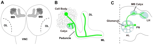
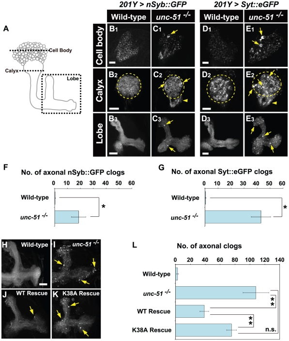
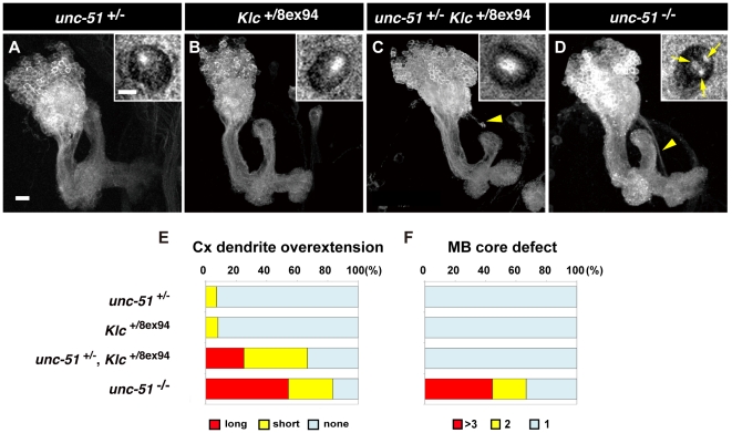
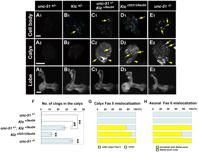
Methodology/principal findings: In this work, we have investigated the functional significance of Unc-51-mediated vesicle transport in the development of complex brain structures in Drosophila. We show that Unc-51 preferentially accumulates in newly elongating axons of the mushroom body, a center of olfactory learning in flies. Mutations in unc-51 cause disintegration of the core of the developing mushroom body, with mislocalization of Fasciclin II (Fas II), an IgG-family cell adhesion molecule important for axonal guidance and fasciculation. In unc-51 mutants, Fas II accumulates in the cell bodies, calyx, and the proximal peduncle. Furthermore, we show that mutations in unc-51 cause aberrant overshooting of dendrites in the mushroom body and the antennal lobe. Loss of unc-51 function leads to marked accumulation of Rab5 and Golgi components, whereas the localization of dendrite-specific proteins, such as Down syndrome cell adhesion molecule (DSCAM) and No distributive disjunction (Nod), remains unaltered. Genetic analyses of kinesin light chain (Klc) and unc-51 double heterozygotes suggest the importance of kinesin-mediated membrane transport for axonal and dendritic development. Moreover, our data demonstrate that loss of Klc activity causes similar axonal and dendritic defects in mushroom body neurons, recapitulating the salient feature of the developmental abnormalities caused by unc-51 mutations.

Conclusions/significance: Unc-51 plays pivotal roles in the axonal and dendritic development of the Drosophila brain. Unc-51-mediated membrane vesicle transport is important in targeted localization of guidance molecules and organelles that regulate elongation and compartmentalization of developing neurons.


Conflict of interest statement

Competing Interests: The authors have declared that no competing interests exist.

Figures

Figure 1
Figure 1. Schematic representations of the larval MB and the adult olfactory PNs.
(A) The Drosophila larval brain. MB, mushroom body; OL, optic lobe primordium; VNC, ventral nerve cord. (B) Structure of the larval MB. The larval MB consists of a single set of dorsal and medial lobes. Each of the MB neurons (highlighted in green) branches dendrites in the calyx and extends its axon through the peduncle, which bifurcates into the dorsal lobe (DL) and medial lobe (ML). (C) The olfactory network in the adult Drosophila brain. The antennal lobe (AL) is the first odor relay station for olfactory information in the fly brain, and consists of approximately 50 glomeruli that are identifiable as discrete neuropil groups. Dendrites of projection neurons (PNs) make specific connections in each different glomerulus in the AL. The PN axons convey olfactory information to higher brain centers by targeting the MB and the lateral horn (LH).
Figure 2
Figure 2. Expression of Unc-51 in the larval brain.
(A–E) Late third instar larval brains stained with an anti-Unc-51 antibody (magenta). MB neurons are labeled with UAS-mCD8::GFP (green) driven by OK107-GAL4. (A) Brain hemisphere of a late third instar larva. OL, optic lobe. Arrows indicate the MB core. (B–D) Localization of Unc-51 in the MB cell bodies (B), calyx (C) and the lobe (D). Note that Unc-51 expression is detected in the cell bodies and the core fibers of the lobe (arrow in D) but not in the calyx (demarcated with a dashed circle). DL, dorsal lobe; ML, medial lobe. (E) Peduncle section showing the localization of Unc-51 in the core fibers, co-stained with phalloidin. UAS-mCD8::GFP (green) was driven by OK107-GAL4, which is expressed in outer layers. (F, G) Peduncle sections showing the localization of kinesin heavy chain (Khc) and kinesin light chain (Klc) in the core fibers (arrows). Note that Fas II is expressed in the outer layers while N-Cadherin (N-Cad) is expressed in the core fibers. (H) Higher magnification image of MB cell bodies. Arrow indicates elevated Unc-51 expression in newly differentiated cells located at the interface of the ganglion mother cells (GMCs) and the Dachshund (DAC)-positive postmitotic neurons. Nb, neuroblast. Cells were labeled with UAS-mCD8::GFP (green) driven by elav-GAL4. Scale bars: (A–D) 50 µm; (E–H) 10 µm.
Figure 3
Figure 3. Loss of unc-51 causes axonal transport defects in MB neurons.
(A) Schematic representation of the larval MB. Dashed lines and box indicate the positions of the cross-sections presented in B–E. (B–E) Localization of n-Syb and Syt1 in the larval MB. Transport of n-Syb and Syt1 was monitored with green fluorescent protein (GFP) fusion constructs expressed in MB neurons using the 201Y-GAL4 driver. Note the accumulations of both markers in cell bodies (C1, E1; arrows), the calyx (C2, E2; arrows) and the lobes (C3, E3; arrows) in unc-51 25/25 mutant MBs. Dashed circles demarcate the calyx. Arrowheads in C2 and E2 indicate aberrant vesicle accumulation in the proximal peduncle. (F, G) Number of axonal clogs. Clog accumulations in the lobes were counted. (F) nSyb::GFP. (G) Syt::eGFP. (H–L) Unc-51 kinase activity is required for axonal transport in MB neurons. (H, I) mCD8::GFP distribution in wild type and unc-51 25/25 mutant MBs. Note the aberrant aggregates in the mutant lobes (arrows in I). (J, K) Genetic rescue of the axonal transport defect in unc-51 mutant by an unc-51 transgene. The axonal aggregation phenotype in unc-51 25/25 mutant was rescued by wild-type unc-51 (J) but not by the kinase-deficient (K38A) mutant, unc-51 K38A (K). UAS-mCD8::GFP and UAS-unc-51 were driven by elav-GAL4. Rescue genotypes: (J) w; elav-GAL4, UAS-mCD8::GFP, UAS-unc-51 WT; unc-51 25/25 and (K) w; elav-GAL4, UAS-mCD8::GFP, UAS-unc-51 K38A; unc-51 25/25. (L) The number of puncta observed in the lobe (mean ± SEM) was plotted for each genotype. **P<0.01 by Student's t test. n.s., not significant (P = 0.125). Scale bars, 10 µm.
Figure 4
Figure 4. unc-51 is required for fasciculation of MB axons.
(A, B) Peduncle sections of late third instar MBs stained with anti-N-Cadherin (green), Fas II (red) and phalloidin (blue). (A) Wild type. (B) unc-51 3/3 mutant. Note the duplicate cores of the mutant. Whereas Fas II is expressed only in mature fibers surrounding the core in the wild type, in the mutant it is aberrantly expressed in a subset of the core fibers (arrow). (C–G) Peduncle sections showing that Unc-51 kinase activity is required for the fasciculation of core fibers. Core fibers were visualized with anti-N-Cadherin. (C) Wild type. (D) unc-51 25/25 mutant. The core defect was rescued by pan-neuronal expression of the unc-51 WT transgene (E) but not by the kinase deficient (K38A) mutant (unc-51 K38A) (F). Genotypes: (E) w; elav-GAL4, UAS-mCD8::GFP, UAS-unc-51 WT; unc-51 25/25 and (F) w; elav-GAL4, UAS-mCD8::GFP, UAS-unc-51 K38A; unc-51 25/25. (G) Number of cores in the peduncle sections. Sixty-seven percent of the unc-51 25/25 mutant MBs had multiple core layers. The core defect was rescued by the unc-51 WT but not the unc-51 K38A transgene. Sample sizes: n = 16 for wild type, n = 18 for unc-51 25/25, n = 6 for unc-51 WT rescue and n = 8 for unc-51 K38A rescue. (H, I) Cell autonomous activity of unc-51 is required for the fasciculation of MB axons. Wild type (H) and unc-51 3/3 mutant (I) neuroblast clones. Mutant clones have wild type-like dorsal lobe (DL) and medial lobe (ML), but exhibit de-fasciculation of the peduncular axons (arrow). (J, K) Cross-sections of the wild-type (J) and mutant (K) peduncles. Clones were induced by a heat shock at the early first instar stage and labeled with mCD8::GFP (green) driven by elav c155. Mature axons were stained with anti-Fas II (magenta). (L) Quantification of the number of peduncular axons in MARCM clones. While only 13% of the wild-type clones (n = 8) exhibited separate bundles, 44% of the unc-51−/− clones (n = 16) demonstrated split fascicles. Scale bars, 10 µm.
Figure 5
Figure 5. Loss of unc-51 causes dendritic defects in MBs.
(A, B) MBs in the wild-type and unc-51 25/25 mutant larvae at the late third instar stage. MBs were visualized with mCD8::GFP driven by 201Y. (C) Quantification of the dendrite overextension phenotype in wild-type and unc-51 mutant larvae. While 14% of the wild-type MBs (n = 21) exhibited short overextensions, 83% of the unc-51 −/− MBs (n = 24) exhibited short or long overextensions (shorter or longer than the diameter of the calyx, respectively). (D, E) Analyses of the dendritic phenotype using MARCM clones. Clones were induced by a heat shock at the early first instar stage. MB neurons were labeled with UAS-mCD8::GFP driven by elav c155. (D) Wild-type neuroblast clone. (E) unc-51 3/3 mutant neuroblast clone. (F) Quantification of the dendrite overextension phenotype in MARCM clones. While only 6% of the wild-type clones (n = 17) showed overextensions, all of the unc-51 3/3 clones (n = 23) exhibited short or long overextensions (shorter or longer than the diameter of the calyx, respectively). (G–J) Dendrite overextension phenotype in unc-51 mutant MBs was rescued by pan-neuronal expression of unc-51 WT but not by the kinase-deficient (K38A) unc-51 K38A transgene. (G) Wild-type. (H) unc-51 25/25 mutant. Rescue genotypes: (I) w; elav-GAL4, UAS-mCD8::GFP, UAS-unc-51 WT; unc-51 25/25 and (J) w; elav-GAL4, UAS-mCD8::GFP, UAS-unc-51 K38A; unc-51 25/25. (K) Quantification of the dendrite overextension phenotype. While none of the wild-type MBs (elav-GAL4, UAS-mCD8::GFP) (n = 6) exhibited calyx overextension, 83% of the unc-51 25/25 mutant MBs (elav-GAL4, UAS-mCD8::GFP; unc-51 25/25) exhibited short or long overextensions (n = 6). The dendritic overextension phenotype was significantly suppressed by the unc-51 WT transgene (w; elav-GAL4, UAS-mCD8::GFP, UAS-unc-51 WT; unc-51 25/25), with 31% of the MBs exhibiting short extensions (n = 16). Expression of unc-51 K38A failed to rescue the phenotype (w; elav-GAL4, UAS-mCD8::GFP, UAS-unc-51 K38A; unc-51 25/25), with 73% of the MBs exhibiting short or long overextensions (n = 22). Note different genetic background from that of the experiments in (A–C). Arrows in (B, E, H and J) indicate overextensions from the calyx. Scale bar, 10 µm.
Figure 6
Figure 6. unc-51 regulates the subcellular localization of Fas II.
(A, B) Localization of Fas II in the wild-type and unc-51 25/25 MBs. In wild type, Fas II (magenta) was localized only to the lobes and the distal peduncle. In unc-51 25/25 mutant MBs, Fas II was mislocalized to the calyx (yellow dashed circles) and the proximal peduncles (arrowheads). MBs were labeled with UAS-mCD8::GFP (green) driven by 201Y-GAL4. (C–H) Localization of Fas II monitored with a YFP fusion construct. Fas II::YFP transgene (green, white) was expressed by the 201Y-GAL4 driver. Counterstained with anti-Dac and anti-N-Cadherin antibodies to visualize the MB neurons (magenta). In wild type (C–E), Fas II::YFP was localized to the lobes and the distal peduncle (not shown), as was the endogenous protein. Loss of unc-51 caused mislocalization of Fas II in the calyx (G). Aberrant Fas II::YFP accumulations (arrows) were observed in the cell bodies (F) and the lobes (H). Note that the internal core was disrupted in the mutant lobes (H). (I) Quantification of the number of axonal clogs in the lobes of the wild type and unc-51−/− mutant clones. (J–Q) Cell autonomous activity of unc-51 is required for intracellular transport of Fas II and Syt1. Wild-type (J–M) and unc-51 3/3 mutant (N–Q) neuroblast clones. Clones were induced by an early first instar heat shock and labeled with UAS-mCD8::GFP driven by elav c155 (green). Fas II was mislocalized in the proximal part of the axons in all the mutant clones (13/13) (arrowheads in M). Most of the mutant clones also exhibited ectopic Syt1 accumulation (10/12) in the proximal part of the MB axons (arrowhead in P). None of the wild-type clones accumulated Fas II (0/9) or Syt1 (0/8) in the corresponding regions. CB, cell bodies; Cx, calyx (indicated by yellow dashed circles). Scale bar, 10 µm.
Figure 7
Figure 7. unc-51 genetically interacts with Klc in dendrite development.
(A–D) Larval MBs of single or double heterozygous mutants. Late third instar stage. MBs were labeled with UAS-mCD8::GFP driven by 201Y-GAL4. Inset panel shows a peduncle cross-section stained with anti-N-Cadherin. Arrowheads indicate overextending dendrites. Arrows in the inset of (D) indicate multiple MB cores in a unc-51 25/25 larva. (E) Quantification of dendritic targeting defects. Sample sizes: unc-51 +/25 (n = 15), Klc+/ 8e×94 (n = 27), Klc+/ 8e×94 unc-51 +/25 (n = 53) and unc-51 25/25 (n = 21). (F) Quantification of MB core defects. Sample sizes: unc-51 +/25 (n = 13), Klc+/ 8e×94 (n = 8), unc-51 +/25 Klc +/8e×94 (n = 12) and unc-51 25/25 (n = 18). Scale bars, 10 µm.
Figure 8
Figure 8. Aberrant Fas II accumulation and mislocalization in unc-51 and Klc mutants.
(A–E) Fas II::YFP distribution patterns in the cell bodies (A1–E1), calyx (A2–E2) and lobes (A3–E3). Fas II::YFP was expressed in MBs using the 201Y-GAL4 driver. Yellow arrows indicate aberrant Fas II clogs. Arrowheads (C2, D2 and E2) indicate Fas II mislocalization in the proximal peduncle. Scale bars, 10 µm. (F) Number of clogs in the calyx. Error bars, mean ± SEM. **P<0.01, Student's t test. Comparisons were also made between Klc c02312/8e×94 and Klc +/8e×94 (**P<0.01) as well as between unc-51 25/25 and unc-51 +/25 (**P<0.01). (G) Quantification of Fas II::YFP mislocalization in the calyx. (H) Quantification of Fas II mislocalization in the proximal peduncle. Sample sizes: unc-51 +/25 (n = 6), Klc +/8e×94 (n = 14), unc-51 +/25 Klc +/8e×94 (n = 4), Klc c02312/8e×94 (n = 8) and unc-51 25/25 (n = 8).

Similar articles

Cited by

References

    1. Guzik BW, Goldstein LS. Microtubule-dependent transport in neurons: steps towards an understanding of regulation, function and dysfunction. Curr Opin Cell Biol. 2004;16:443–450. - PubMed
    1. Sann S, Wang Z, Brown H, Jin Y. Roles of endosomal trafficking in neurite outgrowth and guidance. Trends Cell Biol. 2009;19:317–324. - PubMed
    1. Hirokawa N, Takemura R. Molecular motors in neuronal development, intracellular transport and diseases. Curr Opin Neurobiol. 2004;14:564–573. - PubMed
    1. Barnes AP, Polleux F. Establishment of axon-dendrite polarity in developing neurons. Annu Rev Neurosci. 2009;32:347–381. - PMC - PubMed
    1. Horton AC, Ehlers MD. Neuronal polarity and trafficking. Neuron. 2003;40:277–295. - PubMed

Publication types

LinkOut - more resources