. 2011 May 12;6(5):e19820.
doi: 10.1371/journal.pone.0019820.

Distinct roles of Bcl-2 and Bcl-Xl in the apoptosis of human bone marrow mesenchymal stem cells during differentiation

Affiliations

Distinct roles of Bcl-2 and Bcl-Xl in the apoptosis of human bone marrow mesenchymal stem cells during differentiation

Lisa Oliver et al. PLoS One. .

Abstract

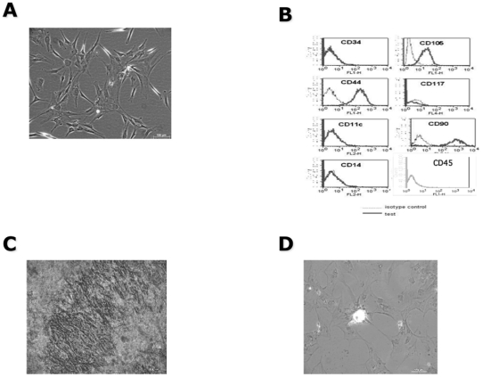
Background: Adult mesenchymal stem cells (MSCs) can be maintained over extended periods of time before activation and differentiation. Little is known about the programs that sustain the survival of these cells.

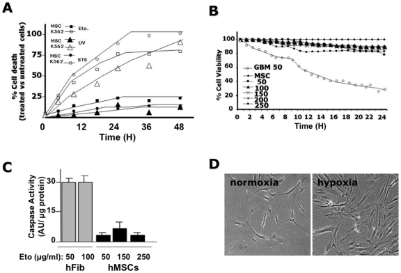
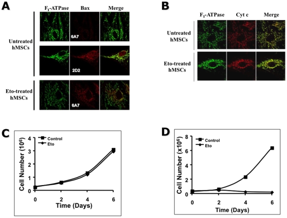
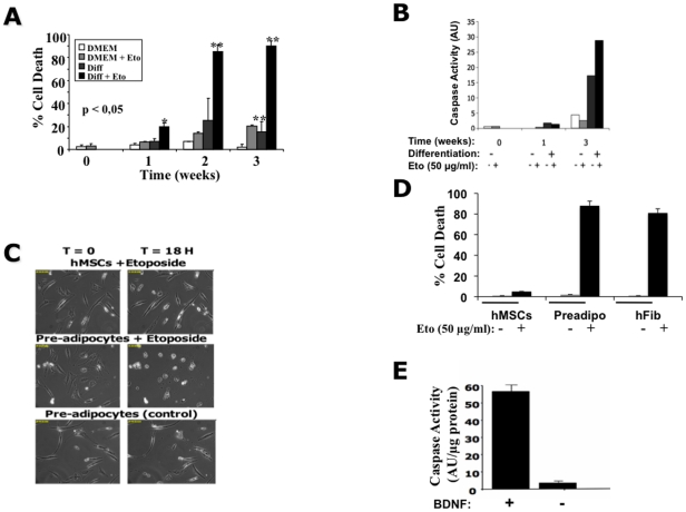
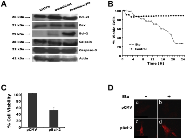
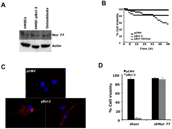
Principal findings: Undifferentiated adult human MSCs (hMSCs) did not undergo apoptosis in response to different cell death inducers. Conversely, the same inducers can readily induce apoptosis when hMSCs are engaged in the early stages of differentiation. The survival of undifferentiated cells is linked to the expression of Bcl-Xl and Bcl-2 in completely opposite ways. Bcl-Xl is expressed at similar levels in undifferentiated and differentiated hMSCs while Bcl-2 is expressed only in differentiated cells. In undifferentiated hMSCs, the down-regulation of Bcl-Xl is associated with an increased sensitivity to apoptosis while the ectopic expression of Bcl-2 induced apoptosis. This apoptosis is linked to the presence of cytoplasmic Nur 77 in undifferentiated hMSCs.

Significance: In hMSCs, the expression of Bcl-2 depends on cellular differentiation and can be either pro- or anti-apoptotic. Bcl-Xl, on the other hand, exhibits an anti-apoptotic activity under all conditions.


Conflict of interest statement

Competing Interests: The authors have declared that no competing interests exist.

Figures

Figure 1
Figure 1. Characterization of in vitro hMSCs.
(A) Cell morphology: Photograph of hMSCs cultured in complete medium as described in materials & methods. Note the fibroblast-like morphology of the cells. (B) Markers (cytometry): Results are represented as FACScan histograms; the gray line corresponds to the isotype control and the solid black line to the specific antibody tested. Data shown are representative of at least three independent experiments. (C, D) hMSCs were cultured for 3 weeks in either osteogenic (C) or adipogenic (D) differentiation media. Matrix mineralization was determined by alkaline phosphatase staining (C) and lipid vacuoles were stained with Oil Red O (D) as described in experimental procedures. The cultures were visualized under an upright Nikon TMS microscope, magnification ×20.
Figure 2
Figure 2. Absence of cell death in hMSCs.
(A) Induction of cell death using staurosporine (STS), UV-irradiation and etoposide (Eto) in K562 cells and hMSCs. The cells were plated at 5×10⁴ cells/ml in 24-well plates. 24 h later the different apoptosis inducing agents were added to the culture medium and the cells were analyzed over 48 h using video-microscopy with an acquisition every 10 min. The number of dead cells was determined at each time point and rounded up for every hour. The results are presented as the percentage of dead cells in treated cultures versus untreated cultures. The number of cells analyzed was about 100 per condition. The results are representative of three independent experiments and of 3 different hMSC cultures. (B) Effect of soluble Fas ligand (sFasL) on hMSCs or GBM primary cultures. Quantification of the viability of hMSCs cultured in the absence or in the presence of increasing concentrations (50, 100, 150, 200 and 250 ng/ml) sFasL and GBM cells cultured in the presence of 50 ng/ml sFasL was determined by time-lapse microscopy over 24 h using 10 min intervals. Cell viability was determined at every acquisition as described in (A. C) Human MSCs or fibroblasts (hfib) were cultured in the absence or in the presence of etoposide (Eto) at the indicated concentrations over 24 h. The cells were then lysed and the caspase activity determined in 20 µg cell extract using Ac-DEVD-AMC as a substrate. The results are expressed as arbitrary units of caspase activity per µg protein. (D) Effect of hypoxia on hMSCs survival. Human MSCs were cultured for 48 h under normoxia (20% O2) or hypoxia using a hypoxia work-station at 3% O2, 5% CO2 in 95% humidified air. Note that experiments shown are representative of hMSCs obtained from at least five different donors.
Figure 3
Figure 3. Absence of release of cyt c from mitochondria despite the activation of Bax in hMSCs.
(A, B) Laser confocal analyses of hMSCs after etoposide treatment. Cells were cultured in the absence (untreated) or in the presence of 50 µg/ml Etoposide (Eto) for 24 h then labeled with polyclonal anti-F1-ATPase (mitochondria: green) and either (A) monoclonal anti-Bax2D2 (2D2: recognizes all forms of Bax: red) and/or anti-Bax6A7 (6A7: recognizes only the activated form of Bax: red) or (B) monoclonal anti-cyt c (red). Laser confocal analyses were done as described in the material and methods section. (C, D) Human MSCs were plated into 6-well plates and 24 h later cultured in the absence or presence of 50 µg/ml etoposide (Eto). 24 h later the cells were either cultured in the presence of complete media (C) or in osteogenic differentiation media (D). At the different times indicated the cells were trypsinized and the number of viable cells determined by trypan blue exclusion.
Figure 4
Figure 4. Sensitivity to cell death was acquired after induction of differentiation.
(A) Human MSCs were cultured in complete or osteogenic differentiation medium for 0 to 3 weeks; in the absence or in the presence of 50 µg/ml etoposide (Eto). The number of viable cells was quantified by Trypan blue exclusion counting a minimum of 200 cells per condition. The data presented represent three independent experiments. (B) The cells treated as in (A) were collected and 10 µg whole cell lysates were assayed for caspase activity using Ac-DEVD-AMC as a substrate. The results are expressed as arbitrary units of caspase activity per µg protein. (C) Induction of apoptosis in hMSCs and adipocytes. Pictomicrographs of adipogenic differentiated hMSCs and hMSCs cultured in the absence or in the presence of 50 µg/ml etoposide (Eto) for 18 h. Images are representative of 4 independent experiments. (D) The number of cell death in hMSCs, preadipocyte (preadipo) or human fibroblast (hfib) cultures treated or not with 50 µg/ml etoposide (Eto) for 24 h was determined by Trypan blue exclusion (Countess), counting about 200 cells under each condition. The data are representative of 3 independent experiments. (E) Human MSCs induced to differentiate along the neuronal pathway (+10 µg/ml BDNF) or not (without BDNF) were cultured in the presence of 50 µg/ml etoposide (Eto) for 24 h before being assayed for caspase activity using Ac-DEVD-AMC as a substrate. The results are expressed as arbitrary units of caspase activity per µg protein.
Figure 5
Figure 5. Expression of key components of the apoptotic machinery in hMSCs.
(A) Western blot analyses of some of the main components of the apoptotic machinery. Total protein extracts were performed and 50 µg protein was analyzed on a 12% SDS-PAGE. Immunodetections were performed with the antibodies cited in the material and methods section. (B) The sensitivity of the hMSCs-shBcl-Xl-501 and hMSCs-shscr to apoptosis was determined by culturing the cells in the absence or in the presence of 50 µg/ml etoposide (Eto). The cells were analyzed over 48 h using video-microscopy with an acquisition every 10 min. The number of dead cells was determined at each time point and rounded up for every hour. The results are presented as the percentage of dead cells in treated cultures versus untreated cultures. The number of cells analyzed was about 100 per condition. The results are representative of three independent experiments. (C) Cells were nucleofected with either pCMV or pBcl-2 and 24 h later cell viability was determined by Trypan blue exclusion. (D) Twenty-four hours after the transfection of hMSCs with pCMV (a, b) or pBcl-2 (c, d); these cells were cultured in the absence (a, c) or presence (b, d) of 50 µg/ml etoposide (Eto) then analyzed for the expression of active caspase 3 (red) by confocal microscopy. The data presented are representative of 2 independent experiments.
Figure 6
Figure 6. Interaction between Bcl-2 and Nur 77.
(A) Western blot analyses of Nur 77 expression in hMSCs, hMSCs transfected with pBcl-2 and osteoblasts. This is a representative immunoblot of 2 different experiments. (B) Cells were transfected with plasmids: pCMV, pCMV containing Bcl-2 (pBcl-2) or Bcl-2 deleted in the amino acids 30–85 (pBcl-2Δloop). 16 h post-transfection the cells were plated in 6-well plated and video-filmed over 48 h with acquisition every 10 min. The number of dead cells was determined at each time point and rounded up for every hour. The results are presented as the percentage of dead cells in treated cultures versus untreated cultures. The number of cells analyzed was minimum 100 per condition. The results are representative of 3 independent experiments. (C) A PLA between Bcl-2 and Nur 77 was performed on hMSCs transfected with pCMV or pBcl-2 as described in the materials and methods section. Fluorescence was detected using the fluorophore Tye-563 that excites at 557 nm and emits at 563 nm. The nuclei were stained with Dapi present in the mounting solution (Prolong® Gold anti-fade). Spots indicated interaction between Nur77 and Bcl-2. These experiments were repeated 3 times, each performed in duplicate. (D) Human MSCs infected with either shNur77 or shscr were then transfected with pCMV or pBcl-2 and then treated with 50 µg/ml etoposide. Note that for all experiments only Nur77-25 was used. Cell viability was determined by Trypan blue exclusion, 24 h after treatments. The results are representative of 3 different experiments done in triplicate.

Similar articles

Cited by

References

    1. He S, Nakada D, Morrison SJ. Mechanisms of stem cell self-renewal. Ann Rev Cell Dev Biol. 2009;25:377–406. - PubMed
    1. Reya T, Morrison SJ, Clarke MF, Weissman IL. Stem cells, cancer, and cancer stem cells. Nature. 2001;414:105–111. - PubMed
    1. Caplan AI. Why are MSCs therapeutic? New data: new insight. J Pathol. 2007;217:318–324. - PMC - PubMed
    1. Uccelli A, Moretta L, Pistoia V. Mesenchymal stem cells in health and disease. Nat Rev Immunol. 2008;8:726–736. - PubMed
    1. Phinney DG, Prockop DJ. Concise review: mesenchymal stem/multipotent stromal cells: the state of transdifferentiation and modes of tissue repair–current views. Stem Cells. 2007;25:2896–2902. - PubMed

Publication types

MeSH terms

Substances