. 2011 May;7(5):e1001340.
doi: 10.1371/journal.ppat.1001340. Epub 2011 May 12.

Quantitative subcellular proteome and secretome profiling of influenza A virus-infected human primary macrophages

Affiliations

Quantitative subcellular proteome and secretome profiling of influenza A virus-infected human primary macrophages

Niina Lietzén et al. PLoS Pathog. 2011 May.

Abstract

Influenza A viruses are important pathogens that cause acute respiratory diseases and annual epidemics in humans. Macrophages recognize influenza A virus infection with their pattern recognition receptors, and are involved in the activation of proper innate immune response. Here, we have used high-throughput subcellular proteomics combined with bioinformatics to provide a global view of host cellular events that are activated in response to influenza A virus infection in human primary macrophages. We show that viral infection regulates the expression and/or subcellular localization of more than one thousand host proteins at early phases of infection. Our data reveals that there are dramatic changes in mitochondrial and nuclear proteomes in response to infection. We show that a rapid cytoplasmic leakage of lysosomal proteins, including cathepsins, followed by their secretion, contributes to inflammasome activation and apoptosis seen in the infected macrophages. Also, our results demonstrate that P2X₇ receptor and src tyrosine kinase activity are essential for inflammasome activation during influenza A virus infection. Finally, we show that influenza A virus infection is associated with robust secretion of different danger-associated molecular patterns (DAMPs) suggesting an important role for DAMPs in host response to influenza A virus infection. In conclusion, our high-throughput quantitative proteomics study provides important new insight into host-response against influenza A virus infection in human primary macrophages.


Conflict of interest statement

The authors have declared that no competing interests exist.

Figures

Figure 1
Figure 1. Quantitative subcellular proteome and secretome analysis of influenza A virus-infected human primary macrophages.
A. Workflow of the experiment. GO = gene ontology. B. Western blot analysis of control and influenza A virus-infected cells from mitochondrial (M), cytoplasmic (C) and nuclear (N) fractions using mitochondrial, cytoplasmic and nuclear markers (voltage-dependent anion-selective channel protein 1 (VDAC1), glyceraldehyde-3-phosphate dehydrogenase (GAPDH) and histone H1, respectively). C. Numbers of reliably quantified and differentially regulated proteins (fold difference ≥1,5 or ≤0,67 for intracellular fractions and ≥3 for secretome) in each subcellular fraction and secretome at different timepoints. D. Gene ontology-based classification of all the proteins identified from intracellular fractions. Mitochondrial, cytoplasmic, nuclear, lysosomal and endoplasmic reticulum (ER) annotations for the identified proteins are shown.
Figure 2
Figure 2. Functional classification of differentially regulated proteins in the nuclear fraction at different timenpoints.
Classification of proteins that are A. upregulated and B. downregulated in the nuclear fractions of influenza A virus-infected macrophages. The numbers of proteins related with each category are shown in brackets.
Figure 3
Figure 3. Influenza A virus infection results in apoptosis and increasing amount of lysosomal proteins in cytoplasm.
A. Examples of differentially expressed apoptosis-related proteins in intracellular fractions. B. Control and influenza A virus-infected macrophages were stained with APOPercentage Apoptosis Assay. Apoptotic cells are stained as purples. C. Lysosomal proteins as well as mitochondrial proteins are overexpressed in the cytoplasmic fraction already at 6 h post-infection. Lysosomal proteins are highlighted as yellow.
Figure 4
Figure 4. Cathepsins are essential for influenza A virus-induced inflammasome activation.
A. Human macrophages were left untreated or infected with influenza A virus for 6, 9, or 12 h, after which concentrated cell culture media was prepared for Western blot analysis with anti-cathepsin B, anti-cathepsin D, anti-caspase-1 Abs. B–C. Human macrophages were infected with influenza A virus in the presence and absence of 40 µM Ca-074 Me, after which the cell culture media was collected and analysed with IL-18 ELISA, and Western blot analysis with anti-IL-18 and caspase-1 specific Abs. D. Human macrophages were infected with influenza A virus in the presence and absence of Ca-074 Me (40 µM). After this total protein lysates were prepared and analyzed by Western blotting with anti-caspase-3 p19/17 and anti-H3N2 specific Abs.
Figure 5
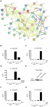
Figure 5. Influenza A virus-induced inflammasome activation is dependent on P2X7 receptor and src tyrosine kinase activity.
A. Protein-protein interaction network of all inflammatory proteins identified from intracellular fractions. B. Human macrophages were infected with influenza A virus for 9 h in the presence and absence of AZ11645373 (1 µM), after which the cell culture media was collected and analyzed with IL-18 ELISA. C. Human macrophages were primed with LPS for 18 h, after which the macrophages were infected with influenza A virus for 9 h in the presence and absence of AZ11645373. After this cell culture supernatants were analyzed with IL-1β ELISA. D. Human macrophages were treated with control siRNA and P2X7 receptor specific siRNAs for 24 h after which the cells were left unstimulated or infected with influenza A virus for 9 h. IL-18 secretion was analyzed from cell culture supernatants with IL-18 ELISA. E. P2X7 receptor expression was analyzed by Western blotting with anti-P2X7 specific Abs. F. Macrophages were infected with influenza A virus for 9 h in the presence and absence of PP2 (5 µM), after which the cell culture media was collected and analyzed with IL-18 ELISA. G. Macrophages were primed with LPS for 18 h, after which the macrophages were left untreated or treated with PP2 and infected with influenza A virus for 9 h. The cell culture supernatants were analyzed with IL-1β ELISA.
Figure 6
Figure 6. High-throughput, quantitative subcellular proteomics provides a global view of host-response in macrophages upon viral infection.

Similar articles

Cited by

References

    1. Akira S, Uematsu S, Takeuchi O. Pathogen recognition and innate immunity. Cell. 2006;124:783–801. - PubMed
    1. Kawai T, Akira S. Innate immune recognition of viral infection. Nat Immunol. 2006;7:131–137. - PubMed
    1. Koyama S, Ishii KJ, Kumar H, Tanimoto T, Coban C, et al. Differential role of TLR- and RLR-signaling in the immune responses to influenza A virus infection and vaccination. J Immunol. 2007;179:4711–4720. - PubMed
    1. Pirhonen J, Sareneva T, Kurimoto M, Julkunen I, Matikainen S. Virus infection activates IL-1β and IL-18 production in human macrophages by a caspase-1-dependent pathway. J Immunol. 1999;162:7322–7329. - PubMed
    1. Pirhonen J, Sareneva T, Julkunen I, Matikainen S. Virus infection induces proteolytic processing of IL-18 in human macrophages via caspase-1 and caspase-3 activation. Eur J Immunol. 2001;31:726–733. - PubMed

Publication types

MeSH terms