. 2011 May;7(5):e1002042.
doi: 10.1371/journal.ppat.1002042. Epub 2011 May 12.

A family of helminth molecules that modulate innate cell responses via molecular mimicry of host antimicrobial peptides

Affiliations

A family of helminth molecules that modulate innate cell responses via molecular mimicry of host antimicrobial peptides

Mark W Robinson et al. PLoS Pathog. 2011 May.

Abstract

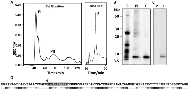
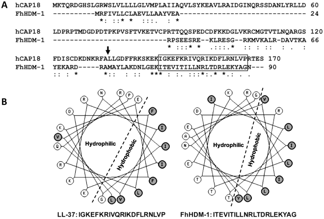
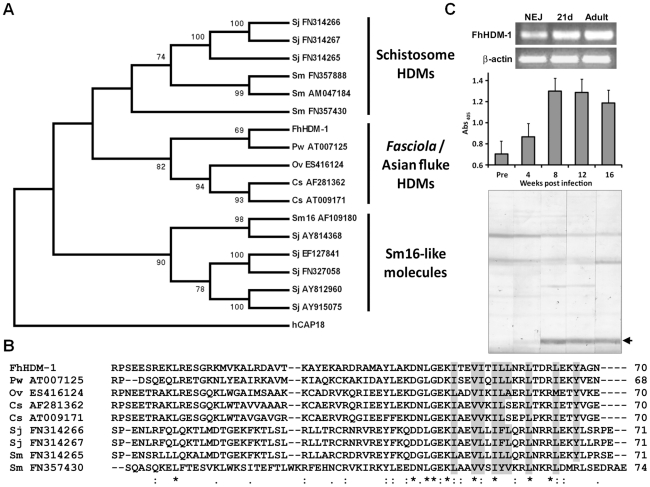
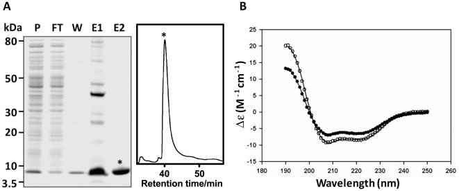
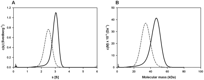
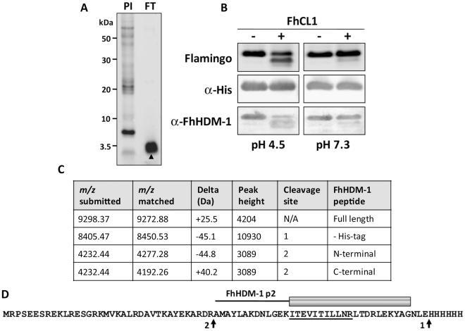
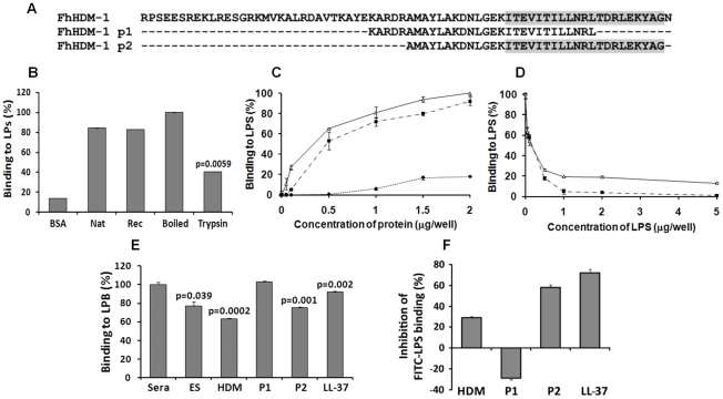
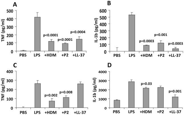
Over the last decade a significant number of studies have highlighted the central role of host antimicrobial (or defence) peptides in modulating the response of innate immune cells to pathogen-associated ligands. In humans, the most widely studied antimicrobial peptide is LL-37, a 37-residue peptide containing an amphipathic helix that is released via proteolytic cleavage of the precursor protein CAP18. Owing to its ability to protect against lethal endotoxaemia and clinically-relevant bacterial infections, LL-37 and its derivatives are seen as attractive candidates for anti-sepsis therapies. We have identified a novel family of molecules secreted by parasitic helminths (helminth defence molecules; HDMs) that exhibit similar biochemical and functional characteristics to human defence peptides, particularly CAP18. The HDM secreted by Fasciola hepatica (FhHDM-1) adopts a predominantly α-helical structure in solution. Processing of FhHDM-1 by F. hepatica cathepsin L1 releases a 34-residue C-terminal fragment containing a conserved amphipathic helix. This is analogous to the proteolytic processing of CAP18 to release LL-37, which modulates innate cell activation by classical toll-like receptor (TLR) ligands such as lipopolysaccharide (LPS). We show that full-length recombinant FhHDM-1 and a peptide analogue of the amphipathic C-terminus bind directly to LPS in a concentration-dependent manner, reducing its interaction with both LPS-binding protein (LBP) and the surface of macrophages. Furthermore, FhHDM-1 and the amphipathic C-terminal peptide protect mice against LPS-induced inflammation by significantly reducing the release of inflammatory mediators from macrophages. We propose that HDMs, by mimicking the function of host defence peptides, represent a novel family of innate cell modulators with therapeutic potential in anti-sepsis treatments and prevention of inflammation.


Conflict of interest statement

The authors have declared that no competing interests exist.

Figures

Figure 1
Figure 1. Identification and characterisation of native FhHDM-1.
(A) Secretory proteins collected from adult F. hepatica following in vitro culture were separated by gel filtration and the resulting high molecular mass (>200 kDa) peak (peak I; PI) was separated further using reverse phase HLPC (RP-HPLC). Fractions collected following gel filtration and RP-HPLC were run on reducing 4–12% Bis-Tris gels (B) and showed that a prominent ∼ 6 kDa protein present in total adult secretory proteins (S) was enriched in PI and purified to homogeneity (>95%) following RP-HPLC (E). (C) Western blot of adult fluke secretions probed with an anti-FhHDM-1 antibody. P, pre-immune sera; T, test bleed. (D) N-terminal sequencing and LC-MS/MS analysis of the native ∼ 6 kDa protein generated peptide sequence information that allowed cloning of the cDNA, termed FhHDM-1. The primary amino acid sequence of FhHDM-1 derived from conceptual translation of the cDNA is shown. The predicted N-terminal signal peptide is shown in italics and the actual N-terminal of the native protein is shown by an arrow. The SEESREKLRE sequence generated by N-terminal sequencing is boxed in grey and a peptide (m/z 642.93; ITEVITILLNR) matched by LC-MS/MS following tryptic digest of the native protein is underlined. Secondary structure predictions using using PSIPRED , shown below the primary sequence, suggest the molecule is predominantly α-helical.
Figure 2
Figure 2. FhHDM-1 is structurally homologous with LL-37.
(A) Primary sequence alignment of FhHDM-1 with the human LL-37 precursor, hCAP18. The LL-37 processing site is arrowed. (B) Helical wheel analysis shows that the conserved C-terminal hydrophobic regions boxed in (A) form amphipathic helices in both molecules.
Figure 3
Figure 3. Phylogenetic relationships of the HDMs.
(A) A bootstrapped (1000 trials) neighbour-joining phylogenetic tree showing the evolutionary relationship of HDM cDNA sequences from medically-important trematode pathogens. Numbers represent bootstrap values (given as percentages) for a particular node, and values greater than 65% are shown. The tree is rooted to human CAP18 (accession number NM_004345). Three major clades are shown corresponding to the Sm16-like molecules, the schistosome HDMs and HDMs from Fasciola and the Asian flukes. (B) Primary sequence alignment of selected members of the HDM clades. Conserved residues that contribute to the hydrophobic face of the amphipathic helix are shaded in grey. (C) Top panel. RT-PCR analysis of FhHDM-1 expression in F. hepatica newly excysted juveniles (NEJ), 21-day immature flukes (21d) and adult worms (Adult). Amplification of constitutively expressed F. hepatica β-actin was performed as a positive control. Samples were separated by agarose gel electrophoresis and stained with ethidium bromide. Bottom panel. Immunogenicity of FhHDM-1 in F. hepatica-infected sheep. Pre-infection sera (Pre) and samples taken 4, 8, 12 and 16 weeks post-infection were analysed by ELISA and Western blot using an anti-FhHDM-1 antibody. Specific antibody responses were detected at week 4 with immunoblot staining stronger at weeks 8 and 12 after infection.
Figure 4
Figure 4. Expression and CD spectroscopy of recombinant FhHDM-1.
(A) The full-length FhHDM-1 cDNA, minus the N-terminal signal peptide, was expressed in E. coli and the His-tagged recombinant was purified from cell lysates using Ni-NTA agarose (Qiagen). P, pre-column; FT, flow-through; W, wash, E1, imidazole eluate. Co-eluting proteins were removed by RP-HLPC resulting in recombinant FhHDM-1 of very high purity (E2). (B) CD spectra of recombinant 0.1 mg/mL−1 FhHDM-1 at pH 7.3. The wavelength scan was performed between 190 and 250 nm. The final spectrum (closed circles in the absence of 30% (v/v) TFE and open circles in the presence of 30% (v/v) TFE) is the average result from three scans measured at 20°C. The CONTINLL algorithm from the CDPro software package produced the best fit (solid lines) against the SP29 protein database with r.m.s.d. values for all samples ≤0.325. FhHDM-1 adopts a near identical solution structure in both native and recombinant form at both pH 4.5 and pH 7.3 (data not shown). The resulting secondary structure proportions are reported in Table S1.
Figure 5
Figure 5. Sedimentation velocity analysis of recombinant FhHDM-1.
(A) Continuous size-distribution analysis, c(s), plotted as a function of sedimentation coefficient for recombinant FhHDM-1 at pH 4.5 (solid line) and pH 7.3 (dashed line). Continuous size-distribution analysis was performed using the program SEDFIT – employing 100 sedimentation coefficients ranging from 0.1 S to 6.0 S and at a confidence level (F-ratio)  = 0.95. (B) Continuous mass, c(M), distribution plotted as a function of molecular mass (kDa) for recombinant FhHDM-1 at pH 4.5 (solid line) and pH 7.3 (dashed line). Continuous mass-distribution analysis was performed using SEDFIT with 100 masses ranging from 1.0 kDa to 80 kDa and at a confidence level (F-ratio) = 0.95.
Figure 6
Figure 6. FhCL1 processes FhHDM-1 at low pH.
(A) Total secretory proteins from adult F. hepatica were concentrated from culture supernatants using a 3 kDa cut-off filter. 10 µg of the flow-through (FT) was analysed on a 4–12% Bi-Tris gel and stained with Flamingo fluorescent protein stain. The FT comprised a single prominent band (∼ 3.5 kDa) that was identified by LC-MS/MS as an FhHDM-1 fragment (high-scoring peptide ITEVITILLNR; m/z 642.93, underlined in D). N-terminal sequencing of this band was unsuccessful. (B) To investigate whether F. hepatica cathepsin L1 (FhCL1) can process FhHDM-1, 50 µg recombinant FhHDM-1 was incubated with 1 µg recombinant FhCL1 in either 0.1 M sodium acetate (pH 4.5) or 0.1 M sodium phosphate (pH 7.3) each containing 1 mM EDTA and 1 mM DTT. Reactions were performed ± FhCL1 for 3 h at 37°C and stopped by the addition of E-64 (10 µM). Samples were analysed on 4–12% Bis-Tris gels and blots were probed with anti-His or anti-FhHDM-1 antibodies. (C) The pH 4.5 reaction in the presence of FhCL1 shown in (B) was analysed by MALDI-TOF MS. The major masses detected correspond to the full length recombinant FhHDM-1 ± the C-terminal His-tag (m/z 9272.88 and 8450.53 respectively) and two fragments (both m/z 4232.44) created by a single cleavage after Arg56 (native peptide numbering). (D) The putative FhHDM-1 cleavage sites are arrowed. Based on this, the synthetic peptide FhHDM-1 p2 was designed (shown as a cartoon above the primary sequence of recombinant FhHDM-1). Whilst trypsinising recombinant FhHDM-1 considerably reduced its interaction with LPS, boiling had no effect.
Figure 7
Figure 7. LPS neutralisation by FhHDM-1.
(A) Alignment of full-length FhHDM-1 with peptide 1 (FhHDM-1 p1) and peptide 2 (FhHDM-1 p2). The conserved C-terminal amphipathic helix is shaded in grey. (B) The ability of native and recombinant FhHDM-1 to bind LPS was investigated by incubating the proteins (2 µg/well) in an LPS-coated (100 ng/well) microtitre plate. Bound proteins were detected by ELISA using rabbit anti-FhHDM-1 as a primary antibody. BSA was used as a baseline control. Whilst trypsinising the recombinant FhHDM-1 significantly reduced the LPS interaction, boiling had no effect. (C) The ability of recombinant FhHDM-1 (Δ), FhHDM-1 p1 (•) or FhHDM-1 p2 (▪) to bind to LPS was investigated by incubating a range of concentrations of proteins (0.02–2 µg/well) in this assay. (D) FhHDM-1 or derived peptides (0.1 µg) were incubated in the presence of LPS (0.05-5 µg/well) and bound peptides measured as described above. Binding of peptides to the LPS-immobilised plates was expressed as a percentage of that measured for 2 µg (for panel C) or 0.1 µg (for panel D) of FhHDM-1. Data are the means ± SD from three separate experiments. (E) FhHDM-1 and FhHDM-1 p2 but not FhHDM-1 p1 reduced the interaction between LPS and LBP as effectively as LL-37. LPS-coated microtitre plates were incubated with 5 µg/well of F. hepatica ES, LL-37, FhHDM-1 or derived peptides for 1 h prior to the addition of 10% human sera in PBS. Interaction of LBP with LPS was measured by ELISA using an anti-LBP primary antibody and expressed as a percentage of that detected for 10% sera in the absence of added peptides. Data are the mean ± SD of three separate experiments. Statistical significance was calculated using the student t-test and represent a comparison to the binding of 10% sera to immobilised LPS. (F) Binding of FITC-conjugated LPS to RAW264.7 cells was inhibited by LL-37, FhHDM-1 and peptides. RAW264.7 cells (5×10⁵cells/ml) were incubated with 100 ng/ml of FITC-conjugated LPS in the presence of FhHDM-1, FhHDM-1 p1, FhHDM-1 p2 and LL-37 (5 µg/ml) in RPMI 1640 containing 10% FBS for 20 min at 4°C. The binding of FITC-LPS was analysed by flow cytometry. Values represent percentage inhibition of FITC-LPS binding compared to cells in the absence of peptides. Data are the mean fluorescence ± SD of three independent experiments.
Figure 8
Figure 8. FhHDM-1 protects mice from LPS-induced inflammation.
(A) BALB/c mice were injected intra-peritoneally with 1 µg of LPS alone or combined with 1 µg of FhHDM-1, FhHDM-1 p2 or LL-37. Two hours later, sera was collected and serum levels of TNF and (B) IL-1β measured by ELISA. (C) Peritoneal macrophages were isolated, cultured unstimulated in media overnight and then levels of TNF and (D) IL-1β in the culture measured by ELISA. Data are the mean ± SD of six mice in each group. Statistical significance represents a comparison to the levels of cytokines secreted by mice given LPS only.

Similar articles

Cited by

References

    1. Medzhitov R. Recognition of microorganisms and activation of the immune response. Nature. 2007;449:819–826. - PubMed
    1. Castellheim A, Brekke OL, Espevik T, Harboe M, Mollnes TE. Innate immune responses to danger signals in systemic inflammatory response syndrome and sepsis. Scand J Immunol. 2009;69:479–491. - PubMed
    1. Giuliani A, Pirri G, Rinaldi AC. Antimicrobial peptides: the LPS connection. Methods Mol Biol. 2010;618:137–154. - PubMed
    1. Hirsch T, Metzig M, Niederbichler A, Steinau HU, Eriksson E, et al. Role of host defense peptides of the innate immune response in sepsis. Shock. 2008;30:117–126. - PubMed
    1. Doss M, White MR, Tecle T, Hartshorn KL. Human defensins and LL-37 in mucosal immunity. J Leukoc Biol. 2010;87:79–92. - PMC - PubMed

Publication types

MeSH terms