. 2011 May;7(5):e1002040.
doi: 10.1371/journal.ppat.1002040. Epub 2011 May 12.

Human neutrophil clearance of bacterial pathogens triggers anti-microbial γδ T cell responses in early infection

Affiliations

Human neutrophil clearance of bacterial pathogens triggers anti-microbial γδ T cell responses in early infection

Martin S Davey et al. PLoS Pathog. 2011 May.

Abstract

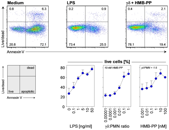
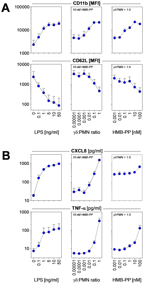
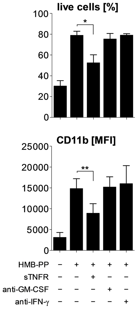
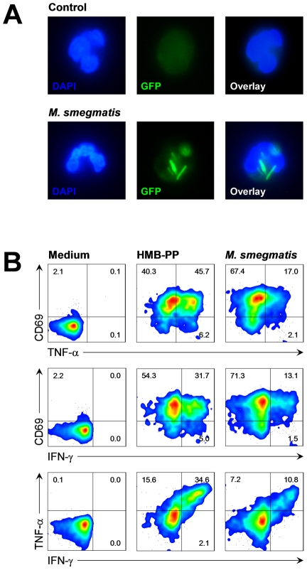
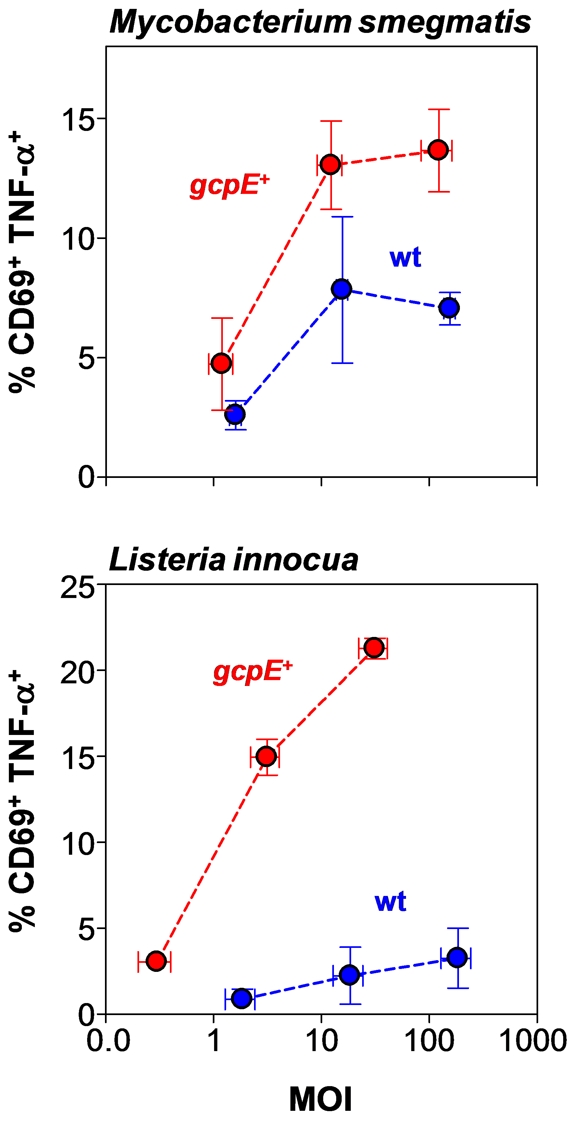
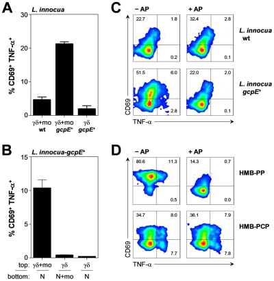
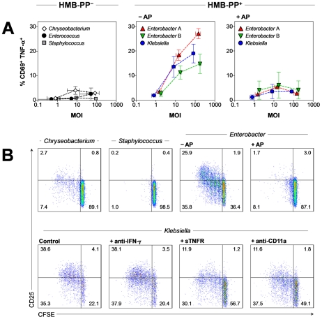
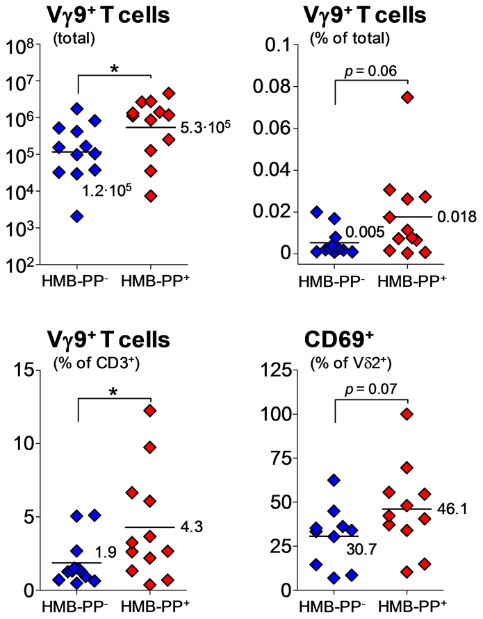
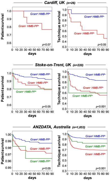
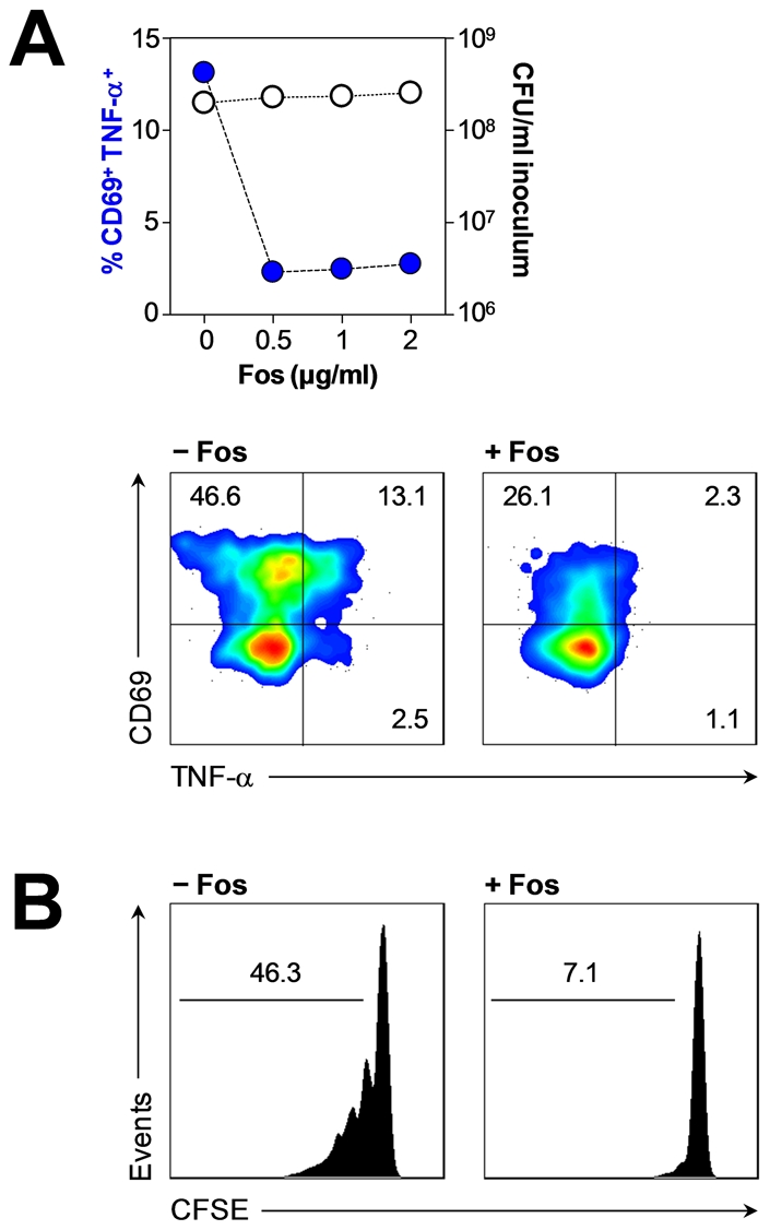
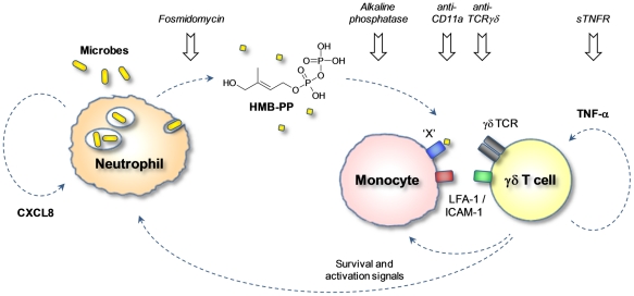
Human blood Vγ9/Vδ2 T cells, monocytes and neutrophils share a responsiveness toward inflammatory chemokines and are rapidly recruited to sites of infection. Studying their interaction in vitro and relating these findings to in vivo observations in patients may therefore provide crucial insight into inflammatory events. Our present data demonstrate that Vγ9/Vδ2 T cells provide potent survival signals resulting in neutrophil activation and the release of the neutrophil chemoattractant CXCL8 (IL-8). In turn, Vγ9/Vδ2 T cells readily respond to neutrophils harboring phagocytosed bacteria, as evidenced by expression of CD69, interferon (IFN)-γ and tumor necrosis factor (TNF)-α. This response is dependent on the ability of these bacteria to produce the microbial metabolite (E)-4-hydroxy-3-methyl-but-2-enyl pyrophosphate (HMB-PP), requires cell-cell contact of Vγ9/Vδ2 T cells with accessory monocytes through lymphocyte function-associated antigen-1 (LFA-1), and results in a TNF-α dependent proliferation of Vγ9/Vδ2 T cells. The antibiotic fosmidomycin, which targets the HMB-PP biosynthesis pathway, not only has a direct antibacterial effect on most HMB-PP producing bacteria but also possesses rapid anti-inflammatory properties by inhibiting γδ T cell responses in vitro. Patients with acute peritoneal-dialysis (PD)-associated bacterial peritonitis--characterized by an excessive influx of neutrophils and monocytes into the peritoneal cavity--show a selective activation of local Vγ9/Vδ2 T cells by HMB-PP producing but not by HMB-PP deficient bacterial pathogens. The γδ T cell-driven perpetuation of inflammatory responses during acute peritonitis is associated with elevated peritoneal levels of γδ T cells and TNF-α and detrimental clinical outcomes in infections caused by HMB-PP positive microorganisms. Taken together, our findings indicate a direct link between invading pathogens, neutrophils, monocytes and microbe-responsive γδ T cells in early infection and suggest novel diagnostic and therapeutic approaches.


Conflict of interest statement

The authors have declared that no competing interests exist.

Figures

Figure 1
Figure 1. HMB-PP stimulated Vγ9/Vδ2 T cells induce neutrophil survival.
Dose-dependent survival of neutrophils incubated with LPS, and of neutrophils (PMN) co-cultured with Vγ9/Vδ2 T cells at various ratios in the presence of HMB-PP at different concentrations. Neutrophils were analyzed after 20 hours in culture; dot plots depict representative annexin-V and fixable live/dead stainings for live CD3 CD15+ neutrophils cultured under the indicated conditions. Data shown are mean percentages (%) + SEM of live cells from independent experiments using three different donors.
Figure 2
Figure 2. HMB-PP stimulated Vγ9/Vδ2 T cells induce neutrophil activation and production of pro-inflammatory mediators.
(A) Dose-dependent up-regulation of CD11b and shedding of CD62L by neutrophils incubated with LPS as positive control, and of neutrophils co-cultured with Vγ9/Vδ2 T cells at various ratios in the presence of HMB-PP at different concentrations. Neutrophils were analyzed after 20 hours in culture; histograms depict representative CD11b and CD62L stainings for live CD3 CD15+ neutrophils cultured under the indicated conditions. Data shown are mean fluorescence intensities (MFI) + SEM from independent experiments using three different donors. (B) Dose-dependent secretion of CXCL8 and TNF-α into the culture supernatant of neutrophils incubated with LPS as positive control, and of neutrophils co-cultured with Vγ9/Vδ2 T cells at various ratios in the presence of HMB-PP at different concentrations. Supernatants were analyzed after 20 hours by ELISA. Data shown are mean levels (pg/ml) + SEM from independent experiments using three different donors.
Figure 3
Figure 3. HMB-PP stimulated Vγ9/Vδ2 T cells induce neutrophil survival and activation through TNF-α.
Neutrophils were co-cultured with Vγ9/Vδ2 T cells for 20 hours at a ratio of 5∶1, in the absence or presence of 10 nM HMB-PP. Soluble cytokines were blocked by the addition of sTNFR, anti-GM-CSF or anti-IFN-γ. Data shown are mean values + SEM for neutrophil survival and expression of CD11b by live neutrophils, as determined in independent experiments using three different donors.
Figure 4
Figure 4. Vγ9/Vδ2 T cells respond to bacteria upon phagocytosis by neutrophils.
(A) Resting neutrophils or neutrophils after phagocytosis of M. smegmatis-gfp + stably expressing GFP, at a multiplicity of infection (MOI) of 10. Cells were counter-stained with DAPI and imaged by fluorescence microscopy. Data shown are representative from independent experiments using two different donors. (B) Activation of Vγ9/Vδ2 T cells co-cultured for 20 hours with neutrophils in the absence (medium) or in the presence of 10 nM HMB-PP, or co-cultured with neutrophils harboring M. smegmatis-gcpE + overexpressing HMB-PP synthase. Data shown are representative from independent experiments using three different donors.
Figure 5
Figure 5. Vγ9/Vδ2 T cells show enhanced responses to phagocytosed bacteria that overproduce HMB-PP.
Dose-dependent activation of Vγ9/Vδ2 T cells by neutrophils harboring genetically engineered transfectants of M. smegmatis (upper panel) or L. innocua (lower panel), in which HMB-PP accumulates intracellularly, compared to the parental wildtype strains (wt). Data shown are mean frequencies of CD69+ TNF-α+ Vγ9/Vδ2 T cells ± SEM after 20 hours in culture, as determined in independent experiments using three different donors. Error bars for MOIs depict mean values ± SEM for the true colony-forming unit (CFU) counts of the bacterial inocula used.
Figure 6
Figure 6. Vγ9/Vδ2 T cells respond, in a monocyte-dependent manner, to phosphatase-sensitive molecules released from phagocytosed bacteria.
(A) Activation of Vγ9/Vδ2 T cells by neutrophils harboring L. innocua wt or L. innocua-gcpE + bacteria, in the presence (γδ-mo) or absence (γδ) of monocytes. Data shown are mean frequencies of CD69+ TNF-α+ Vγ9/Vδ2 T cells + SEM after 20 hours in culture, as determined in independent experiments using three different donors. (B) Activation of Vγ9/Vδ2 T cells in the top chamber of a transwell plate separated from neutrophils (N) harboring L. innocua-gcpE + in the bottom chamber, in the presence or absence of monocytes (mo). Data shown are mean frequencies of CD69+ TNF-α+ Vγ9/Vδ2 T cells + SEM after 20 hours in culture, as determined in independent experiments using three different donors. (C) Activation of Vγ9/Vδ2 T cells co-cultured for 20 hours with monocytes in the presence of supernatants from neutrophils harboring L. innocua wt or L. innocua-gcpE + bacteria that had been pretreated or not with alkaline phosphatase (AP). Data shown are representative from independent experiments using two different donors. (D) Activation of Vγ9/Vδ2 T cells co-cultured for 20 hours with monocytes in the presence of 1 nM HMB-PP or 100 µM HMB-PCP pretreated or not with alkaline phosphatase (AP). Data shown are representative from independent experiments using two different donors.
Figure 7
Figure 7. Vγ9/Vδ2 T cells respond to HMB-PP producing Gram+ and Gram bacteria but not to HMB-PP deficient bacteria.
(A) Dose-dependent activation of Vγ9/Vδ2 T cells by neutrophils harboring clinical isolates of a range of different bacteria: Gram HMB-PP+, Enterobacter cloacae (two different isolates tested, A and B) and Klebsiella pneumoniae; Gram HMB-PP, Chryseobacterium indologenes; and Gram+ HMB-PP, Enterococcus faecalis and Staphylococcus aureus; in the presence or absence of alkaline phosphatase (AP). Data shown are mean frequencies of CD69+ TNF-α+ Vγ9/Vδ2 T cells ± SEM after 20 hours in culture, as determined in independent experiments using 3–5 donors. Error bars for MOIs depict mean values ± SEM for the true CFU counts of the bacterial inocula used. (B) Proliferation after 5 days in culture and CD25 expression of CFSE-labeled Vγ9/Vδ2 T cells in response to supernatants from neutrophils harboring clinical isolates of the indicated bacteria, in the presence or absence of alkaline phosphatase (AP), soluble TNF-α receptor (sTNFR) or blocking antibodies against IFN-γ or CD11a. Data shown are representative of independent experiments using cells from at least two different donors.
Figure 8
Figure 8. Vγ9/Vδ2 T cell numbers and CD69 expression are elevated in acute peritonitis caused by HMB-PP bacteria.
Total numbers and frequencies of Vγ9/Vδ2 T cells (% of total cells and % of all CD3+ T cells), and expression of CD69 on peritoneal Vγ9/Vδ2 T cells in patients with PD-associated peritonitis on day 1 (the day of hospital admission with a cloudy effluent, i.e. before commencement of antibiotic therapy), depending on whether or not the causative pathogen was capable of producing HMB-PP.
Figure 9
Figure 9. Episodes of peritonitis caused by HMB-PP producing bacteria are associated with poor clinical outcome.
Cumulative patient survival (left) and cumulative technique survival (right) of patients with acute bacterial peritonitis, grouped into infections with Gram+ HMB-PP (blue), Gram+ HMB-PP+ (green) or Gram HMB-PP+ pathogens (red); episodes caused by Gram HMB-PP pathogens were not recorded in the patient databases. Top, PD patients admitted at the University Hospital of Wales, Cardiff, with acute peritonitis (day 1, i.e. first presentation with a cloudy bag). Middle, Australian PD patients from the ANZDATA registry with first-time peritonitis. Bottom, PD patients with first-time peritonitis treated at the University Hospital of North Staffordshire, Stoke-on-Trent, UK. Comparisons were made using log-rank tests.
Figure 10
Figure 10. Abrogation of HMB-PP dependent γδ T cell responses by fosmidomycin.
(A) Activation of Vγ9/Vδ2 T cells co-cultured for 20 hours with neutrophils harboring Enterobacter cloacae pre-treated or not for 1 hour with the indicated concentrations of fosmidomycin. The minimum inhibitory concentration (MIC) of fosmidomycin for this bacteria strain was 1 µg/ml, as determined by microbroth dilution (Table S1 in Text S1). Data shown are frequencies of CD69+ TNF-α+ Vγ9/Vδ2 T cells after 20 hours in culture, alongside CFU counts of the original bacterial inocula after 1 hour treatment with fosmidomycin, representative of independent experiments using cells from two different donors. FACS plots show typical Vγ9/Vδ2 T cell responses, representative of independent experiments using cells from three different donors. (B) Proliferation after 5 days in culture of CFSE-labeled Vγ9/Vδ2 T cells in response to supernatants from neutrophils harboring Enterobacter cloacae pre-treated or not for 1 hour with 0.25 µg/ml fosmidomycin. Data shown are representative of independent experiments using cells from two different donors.
Figure 11
Figure 11. HMB-PP dependent interaction between γδ T cells, neutrophils and monocytes in acute microbial infection.
Local secretion of inflammatory chemokines leads to extravasation of neutrophils, monocytes and γδ T cells toward the site of infection. Upon phagocytosis of invading microbes, neutrophils release traces of HMB-PP into the microenvironment where it becomes ‘visible’ to γδ T cells. γδ T cells recognize HMB-PP in the context of a yet unidentified presenting molecule ‘X’ and contact-dependent signals provided by monocytes. Crosstalk between the three different cell types triggers the production of pro-inflammatory cytokines such as TNF-α, which drives local γδ T cell expansion, and chemokines such as CXCL8, which recruits further neutrophils to the site of infection. Activated γδ T cells also provide survival and activation signals such as TNF-α for newly arriving neutrophils and monocytes. This γδ T cell-driven inflammatory reaction can be interrupted at various check-points as demonstrated in the present study.

Similar articles

Cited by

References

    1. Nathan N. Neutrophils and immunity: challenges and opportunities. Nat Rev Immunol. 2006;6:173–182. - PubMed
    1. Müller I, Munder M, Kropf P, Hänsch GM. Polymorphonuclear neutrophils and T lymphocytes: strange bedfellows or brothers in arms? Trends Immunol. 2009;30:522–530. - PubMed
    1. Weaver CT, Hatton RD, Mangan PR, Harrington LE. IL-17 family cytokines and the expanding diversity of effector T cell lineages. Annu Rev Immunol. 2007;25:821–852. - PubMed
    1. Sallusto F, Lanzavecchia A. Heterogeneity of CD4+ memory T cells: functional modules for tailored immunity. Eur J Immunol. 2009;39:2076–2082. - PubMed
    1. Annunziato F, Romagnani S. Heterogeneity of human effector CD4+ T cells. Arthritis Res Ther. 2009;11:257. - PMC - PubMed

Publication types

MeSH terms

Substances