Comparative Study
. 2012 Jun;34(3):669-79.
doi: 10.1007/s11357-011-9264-y. Epub 2011 May 18.

Age associated low mitochondrial biogenesis may be explained by lack of response of PGC-1α to exercise training

Affiliations
Comparative Study

Age associated low mitochondrial biogenesis may be explained by lack of response of PGC-1α to exercise training

Frederic Derbré et al. Age (Dordr). 2012 Jun.

Abstract

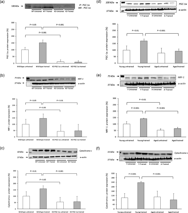
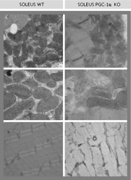
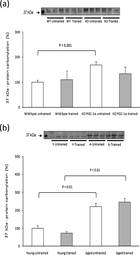
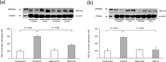
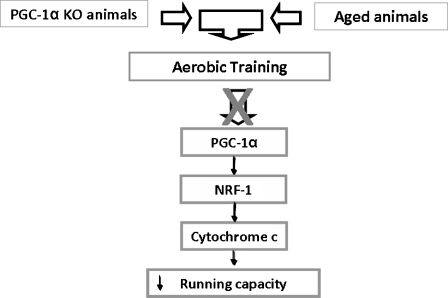
Low mitochondriogenesis is critical to explain loss of muscle function in aging and in the development of frailty. The aim of this work was to explain the mechanism by which mitochondriogenesis is decreased in aging and to determine to which extent it may be prevented by exercise training. We used aged rats and compared them with peroxisome proliferator-activated receptor-γ coactivator-1α deleted mice (PGC-1α KO). PGC-1α KO mice showed a significant decrease in the mitochondriogenic pathway in muscle. In aged rats, we found a loss of exercise-induced expression of PGC-1α, nuclear respiratory factor-1 (NRF-1), and of cytochrome C. Thus muscle mitochondriogenesis, which is activated by exercise training in young animals, is not in aged or PGC-1α KO ones. Other stimuli to increase PGC-1α synthesis apart from exercise training, namely cold induction or thyroid hormone treatment, were effective in young rats but not in aged ones. To sum up, the low mitochondrial biogenesis associated with aging may be due to the lack of response of PGC-1α to different stimuli. Aged rats behave as PGC-1α KO mice. Results reported here highlight the role of PGC-1α in the loss of mitochondriogenesis associated with aging and point to this important transcriptional coactivator as a target for pharmacological interventions to prevent age-associated sarcopenia.


Figures

Fig. 1
Fig. 1
Exercise-induced activation of the mitochondrial biogenesis pathway in skeletal muscle. Western blotting analysis to detect PGC-1α (a and d), NRF-1 (b and e), and cytochrome C (c and f) in PGC-1α KO mice and aged rats. Twenty-seven male mice were randomly divided into four experimental groups: WT untrained (n = 6), WT trained (n = 8), KO untrained (n = 7), and KO trained (n = 6). Twenty-four male Wistar rats were randomly divided into four experimental groups: young untrained (n = 6), young trained (n = 6), aged untrained (n = 6), and aged trained (n = 6). Representative blots are shown. For the densitometric analysis of the results, values are shown as mean (±SD).The content of α-actin, a housekeeping protein marker in skeletal muscle, was determined in all the experimental groups
Fig. 2
Fig. 2
Electron microscopy analysis of soleus muscle from WT and PGC-1α KO animals. Smaller mitochondria were found in the muscle of PGC-1α KO mice compared to sex- and age-matched WT
Fig. 3
Fig. 3
Muscle protein oxidation in untrained and trained animals. Western blotting analysis to detect carbonylated proteins (MW: 37 kDa). Representative experiments are shown. For the densitometric analysis of the results values are shown as mean (±SD) of a WT untrained (n = 6), WT trained (n = 8), PGC-1α KO untrained (n = 7), and PGC-1α KO-trained animals (n = 6); b young untrained (n = 6), young trained (n = 6), aged untrained (n = 6), and aged-trained animals (n = 6)
Fig. 4
Fig. 4
PGC-1α protein levels in skeletal muscle of animals exposed to cold (4 ± 1°C) or treated with triiodothyronine (0.4 mg × kg−1). Representative experiments are shown. For the densitometric analysis of the results values are shown as mean (±SD) of a young control (n = 4), young exposed to cold (n = 4), aged control (n = 4), and aged-exposed to cold (n = 4); b young control (n = 4), young treated with T3 (n = 4), aged control (n = 4), aged treated with T3 (n = 4). The content of α-actin, a housekeeping protein marker in skeletal muscle, was determined in all the experimental groups
Fig. 5
Fig. 5
PGC-1α is not functional in the aged skeletal muscle and it can be involved in the decrease in mitochondrial biogenesis during aging. Proposed mechanism

References

    1. Anderson RM, Weindruch R. Metabolic reprogramming, caloric restriction and aging. Trends Endocrinol Metab. 2009;21(3):134–141. doi: 10.1016/j.tem.2009.11.005. - DOI - PMC - PubMed
    1. Anderson RM, Barger JL, Edwards MG, Braun KH, O’Connor CE, Prolla TA, Weindruch R. Dynamic regulation of PGC-1alpha localization and turnover implicates mitochondrial adaptation in calorie restriction and the stress response. Aging Cell. 2008;7(1):101–111. doi: 10.1111/j.1474-9726.2007.00357.x. - DOI - PMC - PubMed
    1. Davies KJ, Packer L, Brooks GA. Biochemical adaptation of mitochondria, muscle, and whole-animal respiration to endurance training. Arch Biochem Biophys. 1981;209(2):539–554. doi: 10.1016/0003-9861(81)90312-X. - DOI - PubMed
    1. Davies KJ, Quintanilha AT, Brooks GA, Packer L. Free radicals and tissue damage produced by exercise. Biochem Biophys Res Commun. 1982;107(4):1198–1205. doi: 10.1016/S0006-291X(82)80124-1. - DOI - PubMed
    1. Drew B, Phaneuf S, Dirks A, Selman C, Gredilla R, Lezza A, Barja G, Leeuwenburgh C. Effects of aging and caloric restriction on mitochondrial energy production in gastrocnemius muscle and heart. Am J Physiol Regul Integr Comp Physiol. 2003;284(2):R474–R480. - PubMed

Publication types

MeSH terms

Substances