. 2011 Sep;26(9):2151-60.
doi: 10.1002/jbmr.425.

Connexin43 deficiency reduces the sensitivity of cortical bone to the effects of muscle paralysis

Affiliations

Connexin43 deficiency reduces the sensitivity of cortical bone to the effects of muscle paralysis

Susan K Grimston et al. J Bone Miner Res. 2011 Sep.

Abstract

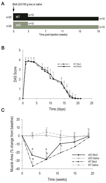
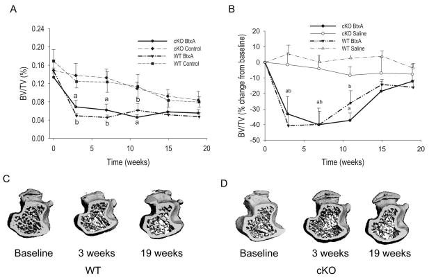
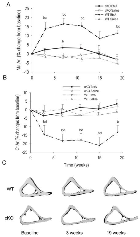
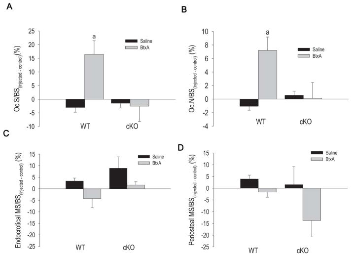
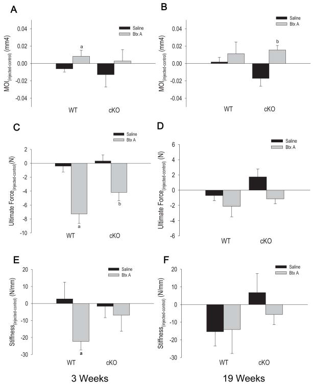
We have shown previously that the effect of mechanical loading on bone depends in part on connexin43 (Cx43). To determine whether Cx43 is also involved in the effect of mechanical unloading, we have used botulinum toxin A (BtxA) to induce reversible muscle paralysis in mice with a conditional deletion of the Cx43 gene in osteoblasts and osteocytes (cKO). BtxA injection in hind limb muscles of wild-type (WT) mice resulted in significant muscle atrophy and rapid loss of trabecular bone. Bone loss reached a nadir of about 40% at 3 weeks after injection, followed by a slow recovery. A similar degree of trabecular bone loss was observed in cKO mice. By contrast, BtxA injection in WT mice significantly increased marrow area and endocortical osteoclast number and decreased cortical thickness and bone strength. These changes did not occur in cKO mice, whose marrow area is larger, osteoclast number higher, and cortical thickness and bone strength lower relative to WT mice in basal conditions. Changes in cortical structure occurring in WT mice had not recovered 19 weeks after BtxA injection despite correction of the early osteoclast activation and a modest increase in periosteal bone formation. Thus BtxA-induced muscle paralysis leads to rapid loss of trabecular bone and to changes in structural and biomechanical properties of cortical bone, neither of which are fully reversed after 19 weeks. Osteoblast/osteocyte Cx43 is involved in the adaptive responses to skeletal unloading selectively in the cortical bone via modulation of osteoclastogenesis on the endocortical surface.


Conflict of interest statement

CONFLICTS OF INTEREST

Roberto Civitelli has a Material Transfer Agreement with Zealand Pharma, Glostrup, Denmark, for the use of gap junction modifying peptides, but receives no honoraria or research funds from Zealand. He receives consultant fees from Novartis and Amgen, grant support from Eli-Lilly and Pfizer; and own stock of Eli-Lilly, Merck and Amgen. None of the other authors have financial conflicts of interest.

Figures

Figure 1
Figure 1
Study design and effect of BtxA injection on muscle function and mass. A. Diagrammatic representation of the study design. A total of 40 mice were used, 20 of each genotype. Twenty mice were sacrificed 3 weeks after intramuscular injection of botulinum toxin A (BtxA) or saline (5 per treatment for each genotype); and 20 were followed up to 19 weeks (5 per treatment for each genotype). The mice followed for 19 weeks had in vivo μCT scans every 4 weeks. B. Changes in muscle function assessed by Digital Abduction Score (DAS) after BtxA injection. Muscle paralysis (DAS=4) and recovery were similar in wild type (WT) and conditional knockout (cKO). C. Change in muscle area measured by μCT in WT and cKO mice followed for 19 weeks. There was a significant effect of treatment on muscle area by 2-way ANOVA (p<0.05); p < 0.05. a: p < 0.05 vs cKO Saline; b: p < 0.05 vs WT Saline.
Figure 2
Figure 2
Effect of botulinum toxin A (BtxA) on trabecular bone in wild type (WT) and Gja1 conditional knockout (cKO) mice. A. Trabecular bone volume (BV/TV) in injected (BtxA) and control limbs of mice followed for 19 weeks. There was a significant decrease of BV/TV in the tibiae in the BtxA injected limbs compared with control tibiae. a: p < 0.05 vs cKO BtxA Control; b: p < 0.05 vs WT BtxA Control. B. Difference in BV/TV between injected (BtxA or saline) and non-injected control limbs, expressed as a percentage of baseline. Following the injection, the difference (imbalance) between injected and non-injected limbs sharply decreased selectively in the BtxA but not in the saline groups of each genotype. There is a progressive though incomplete recovery thereafter. There was a significant effect of treatment and time (p<0.05) but no effect of genotype by 2-way ANOVA. a: p < 0.05 vs cKO Saline; b: p < 0.05 vs WT Saline. C. Micro-CT generated 3-D reconstruction of metaphyses of WT BtxA and (D) tibiae of cKO BtxA injected limbs at different time-points.
Figure 3
Figure 3
Effect of botulinum toxin A (BtxA) on cortical bone in wild type (WT) and Gja1 conditional knockout (cKO). A. Difference in Marrow Area (Ma.Ar.) between injected (BtxA or saline) and non-injected control limbs, expressed as a percentage of baseline. There was significant expansion of the marrow cavity for WT mice beginning 3 weeks post BtxA injection and persisting through the 19 weeks of the study. There was a significant effect of genotype and treatment by 2-way ANOVA (p < 0.05). B. Difference in Cortical Area (Ct.Ar) between injected (BtxA or saline) and non-injected control limbs, expressed as a percentage of baseline. There was a significant reduction in relative cortical area over time for the WT BtxA mice. There was a significant genotype effect in the BtxA group (p<0.05) but not in the Saline mice by 2-way ANOVA (p>0.05). No significant changes in Ct.Ar were observed for the cKO BtxA mice over the 19 weeks of the study. C. Micro-CT generated 3-D reconstruction of diaphyses of WT and cKO taken at baseline, 3 weeks and 19 weeks for each genotype. a: p < 0.05 vs cKO Saline, b: p < 0.05 vs WT Saline, c: p < 0.05 vs cKO BtxA, d: p < 0.05 vs WT Baseline.
Figure 4
Figure 4
The effect of botulinum toxin A (BtxA) on in wild type (WT) and Gja1 conditional knockout (cKO) mice on parameters of bone resorption as determined via TRAP staining for osteoclasts at the mid diaphysis (n=12 per genotype group), and measures of bone formation as determined via calcein labeling. Results are expressed as the absolute difference from the Control limb. A. osteoclast surface/bone surface (OcS/BS) for mice sacrificed 3 weeks after BtxA injection. a: p < 0.05 vs WT Saline. B. Number of osteoclasts per BS (N. Oc./BS) for mice sacrificed 3 weeks after BtxA injection. a: p < 0.05 vs WT Saline. C. Mineralizing surface/bone surface (MS/BS) at the endocortical surface of the mid-diaphysis in mice sacrificed 3 weeks post BtxA injection. D. Mineralizing surface/bone surface (MS/BS) at the periosteal surface of the mid diaphysis. There were no significant differences in terms of mineralizing surface between groups.
Figure 5
Figure 5
Effect of Botulinum toxin A (BtxA) on biomechanical parameters at the mid diaphysis for wild type (WT) and Gja1 conditional knockout (cKO) three weeks post injection and 19 weeks post injection. Results are expressed as the absolute change from the control limb of each group. A. Second area moment of inertia (MOI) values from mice sacrificed 3 weeks post-injection. a: p < 0.05 versus WT Saline. B. Second area moment of inertia (MOI) for WT and cKO, sacrificed 19 weeks post injection. b: p < 0.05 vs WT Saline. C. Ultimate Force (bone strength) data for mice sacrificed 3 weeks after BtxA injection. a: p < 0.05 vs WT Saline. D. Ultimate force (bone strength) data for mice sacrificed 19 weeks post-injection for WT and cKO mice. There were no significant differences in ultimate force between the BtxA and Saline groups at this later time point. E. Bone stiffness results for WT and cKO mice three weeks post BtxA injection. a: p < 0.05 versus WT Saline; F. Bone stiffness results for WT and cKO mice sacrificed after 19 weeks. b: p < 0.05 vs cKO Saline.

References

    1. Lecanda F, Warlow PM, Sheikh S, Furlan F, Steinberg TH, Civitelli R. Connexin43 deficiency causes delayed ossification, craniofacial abnormalities, and osteoblast dysfunction. J Cell Biol. 2000;151(4):931–944. - PMC - PubMed
    1. Paznekas WA, Boyadjiev SA, Shapiro RE, Daniels O, Wollnik B, Keegan CE, Innis JW, Dinulos MB, Christian C, Hannibal MC, Jabs EW. Connexin 43 (GJA1) mutations cause the pleiotropic phenotype of oculodentodigital dysplasia. Am J Hum Genet. 2003;72(2):408–418. - PMC - PubMed
    1. Kjaer KW, Hansen L, Eiberg H, Leicht P, Opitz JM, Tommerup N. Novel Connexin 43 (GJA1) mutation causes oculo-dento-digital dysplasia with curly hair. Am J Med Genet. 2004;127A(2):152–157. - PubMed
    1. Flenniken AM, Osborne LR, Anderson N, Ciliberti N, Fleming C, Gittens JE, Gong XQ, Kelsey LB, Lounsbury C, Moreno L, Nieman BJ, Peterson K, Qu D, Roscoe W, Shao Q, Tong D, Veitch GI, Voronina I, Vukobradovic I, Wood GA, Zhu Y, Zirngibl RA, Aubin JE, Bai D, Bruneau BG, Grynpas M, Henderson JE, Henkelman RM, McKerlie C, Sled JG, Stanford WL, Laird DW, Kidder GM, Adamson SL, Rossant J. A Gja1 missense mutation in a mouse model of oculodentodigital dysplasia. Development. 2005;132(19):4375–4386. - PubMed
    1. Chung DJ, Castro CH, Watkins M, Stains JP, Chung MY, Szejnfeld VL, Willecke K, Theis M, Civitelli R. Low peak bone mass and attenuated anabolic response to parathyroid hormone in mice with an osteoblast-specific deletion of connexin43. J Cell Sci. 2006;119(Pt 20):4187–98. - PubMed

Publication types

MeSH terms