. 2011 Jun;65(6):1711-7.
doi: 10.1002/mrm.22742. Epub 2010 Dec 16.

Novel 16-channel receive coil array for accelerated upper airway MRI at 3 Tesla

Affiliations

Novel 16-channel receive coil array for accelerated upper airway MRI at 3 Tesla

Yoon-Chul Kim et al. Magn Reson Med. 2011 Jun.

Abstract

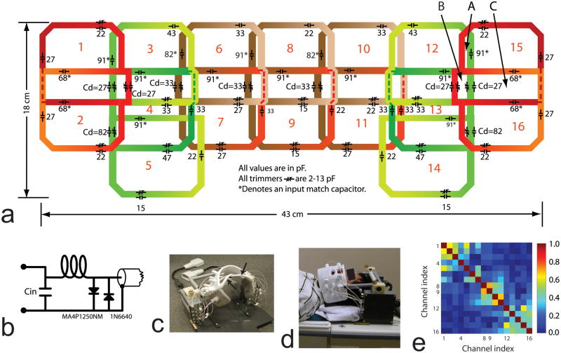
Upper airway MRI can provide a noninvasive assessment of speech and swallowing disorders and sleep apnea. Recent work has demonstrated the value of high-resolution three-dimensional imaging and dynamic two-dimensional imaging and the importance of further improvements in spatio-temporal resolution. The purpose of the study was to describe a novel 16-channel 3 Tesla receive coil that is highly sensitive to the human upper airway and investigate the performance of accelerated upper airway MRI with the coil. In three-dimensional imaging of the upper airway during static posture, 6-fold acceleration is demonstrated using parallel imaging, potentially leading to capturing a whole three-dimensional vocal tract with 1.25 mm isotropic resolution within 9 sec of sustained sound production. Midsagittal spiral parallel imaging of vocal tract dynamics during natural speech production is demonstrated with 2 × 2 mm(2) in-plane spatial and 84 ms temporal resolution.


Figures

Figure 1
Figure 1
16-channel upper airway receive coil array. (a): Coil layout. Individual coil elements are overlapped with their neighboring coil elements to minimize mutual inductive coupling. The two lower coil elements, which correspond to the arrows in (c), extend down the side of the neck region to improve SNR of the lower airway. Channel indices are indicated by the red colored numbers. (b): Input circuit diagram. (c): Photograph of the 16-channel coil. Each coil element is connected to a preamplifier. A pivoting cross bar is supported by the two preamplifier boxes and holds an adjustable cantilever connected to the coil array. The coil array can be held close to the face or folded up and back to permit patient entry and exit. (d): Side view of the 16-channel coil placed on a supine volunteer. (e): Noise correlation matrix from one representative volunteer.
Figure 2
Figure 2
Illustration of coil sensitivity and the upper airway regions of interest (ROIs) used in the evaluation of SNR. Shown is a midsagittal image from Subject 3 obtained using the 16-channel upper airway coil. Note that the image has the most highly localized sensitivity over the upper airway. The eight ROIs identified by red contours are numbered as follows: 1—upper lip, 2—lower lip, 3—front tongue, 4—mid tongue, 5—back tongue, 6—palate, 7—velum, 8—pharyngeal wall.
Figure 3
Figure 3
2D midsagittal image reconstruction using 1D SENSE. The direction of undersampling was A-P. SENSE reconstructed images with a spatial resolution of 1.88 × 1.88 × 2.50 mm3 are compared with respect to reduction factors from 2 to 5 for the (a) Subject 1 and (c) Subject 2. Corresponding g-factor maps are shown for the (b) Subject 1 and (d) Subject 2. The mean and maximum g-factors shown were computed from the ROIs indicated by the red contours of the R = 1 images in (a) and (c).
Figure 4
Figure 4
3D image reconstruction using 2D SENSE. The directions of undersampling were A-P and R-L. Fully sampled 1.25 mm isotropic resolution 3D data set was acquired in open-mouthed position during 54 seconds scan. Retrospective undersampling was performed with a regular undersampling factor of 6, where two-fold acceleration was along R-L direction and three-fold acceleration was along A-P direction. Reformatted midsagittal images are shown for (a) SENSE R = 1, and (c) SENSE R = 6. Eight representative coronal and axial images are shown for (b) SENSE R = 1 and (d) SENSE R = 6. Air-tissue boundaries that are relevant to the vocal tract shape measurement are well defined in all the SENSE R=6 images.
Figure 5
Figure 5
2D image reconstruction of the dynamics of vocal tract shaping using spiral SENSE. (a) Comparison of spiral SENSE images from a single representative frame. For the evaluation of SENSE reconstruction performance under different reduction factors, the frame was chosen from the data acquired when the subject (Subject 1) was stationary. Spatial aliasing was substantially reduced for all the SENSE reconstructed images. The R = 6.8 image exhibited relatively lower image SNR in the posterior regions of the upper airway (e.g., velum and pharyngeal wall). (b) Dynamic frames that represent the subject’s utterance of “ee shop”. The R = 4.2 SENSE reconstruction was used to generate the image frames with its frame update rate of 44 ms. The frame sequence is ordered from the top left to the bottom right.

References

    1. Shellock FG, Schatz CJ, Julien P, Steinberg F, Foo TK, Hopp ML, Westbrook PR. Occlusion and narrowing of the pharyngeal airway in obstructive sleep apnea: evaluation by ultrafast spoiled GRASS MR imaging. Am J Roentgenology. 1992;158:1019–1024. - PubMed
    1. Arens R, McDonough JM, Costarino AT, Mahboubi S, Tayag-Kier CE, Maislin G, Schwab RJ, Pack AI. Magnetic resonance imaging of the upper airway structure of children with obstructive sleep apnea syndrome. Am J Respir Crit Care Med. 2001;164:698–703. - PubMed
    1. Hartl DM, Albiter M, Kolb F, Luboinski B, Sigal R. Morphologic parameters of normal swallowing events using single-shot fast spin echo dynamic MRI. Dysphagia. 2003;18:255–262. - PubMed
    1. Narayanan SS, Alwan AA, Haker K. An articulatory study of fricative consonants using magnetic resonance imaging. J Acoust Soc Am. 1995;98(3):1325–1347.
    1. Kim YC, Narayanan SS, Nayak KS. Accelerated three-dimensional upper airway MRI using compressed sensing. Magn Reson in Med. 2009;61(6):1434–1440. - PMC - PubMed

Publication types