The diverse pathology and kinetics of mass, nonmass, and focus enhancement on MR imaging of the breast
- PMID: 21591007
- PMCID: PMC3098464
- DOI: 10.1002/jmri.22567
The diverse pathology and kinetics of mass, nonmass, and focus enhancement on MR imaging of the breast
Abstract
Purpose: To compare the pathology and kinetic characteristics of breast lesions with focus-, mass-, and nonmass-like enhancement.
Materials and methods: A total of 852 MRI detected breast lesions in 697 patients were selected for an IRB approved review. Patients underwent dynamic contrast enhanced MRI using one pre- and three to six postcontrast T(1)-weighted images. The "type" of enhancement was classified as mass, nonmass, or focus, and kinetic curves quantified by the initial enhancement percentage (E(1)), time to peak enhancement (T(peak)), and signal enhancement ratio (SER). These kinetic parameters were compared between malignant and benign lesions within each morphologic type.
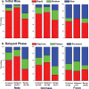
Results: A total of 552 lesions were classified as mass (396 malignant, 156 benign), 261 as nonmass (212 malignant, 49 benign), and 39 as focus (9 malignant, 30 benign). The most common pathology of malignant/benign lesions by morphology: for mass, invasive ductal carcinoma/fibroadenoma; for nonmass, ductal carcinoma in situ (DCIS)/fibrocystic change(FCC); for focus, DCIS/FCC. Benign mass lesions exhibited significantly lower E(1), longer T(peak), and lower SER compared with malignant mass lesions (P < 0.0001). Benign nonmass lesions exhibited only a lower SER compared with malignant nonmass lesions (P < 0.01).
Conclusion: By considering the diverse pathology and kinetic characteristics of different lesion morphologies, diagnostic accuracy may be improved.
Copyright © 2011 Wiley-Liss, Inc.
Figures



Similar articles
-
DCEMRI of breast lesions: is kinetic analysis equally effective for both mass and nonmass-like enhancement?Med Phys. 2008 Jul;35(7):3102-9. doi: 10.1118/1.2936220. Med Phys. 2008. PMID: 18697535 Free PMC article.
-
Nonmass Enhancement Breast Lesions: Diagnostic Performance of Kinetic Assessment on Ultrafast and Standard Dynamic Contrast-Enhanced MRI in Comparison With Morphologic Evaluation.AJR Am J Roentgenol. 2020 Aug;215(2):511-518. doi: 10.2214/AJR.19.21920. Epub 2020 May 26. AJR Am J Roentgenol. 2020. PMID: 32452698
-
Improving the Accuracy of Computer-aided Diagnosis for Breast MR Imaging by Differentiating between Mass and Nonmass Lesions.Radiology. 2016 Mar;278(3):679-88. doi: 10.1148/radiol.2015150241. Epub 2015 Sep 18. Radiology. 2016. PMID: 26383229
-
Breast DCE-MRI: influence of postcontrast timing on automated lesion kinetics assessments and discrimination of benign and malignant lesions.Acad Radiol. 2014 Sep;21(9):1195-203. doi: 10.1016/j.acra.2014.04.013. Epub 2014 Jul 4. Acad Radiol. 2014. PMID: 24998690 Free PMC article.
-
Three-phase dynamic breast magnetic resonance imaging with two-way subtraction.J Comput Assist Tomogr. 2005 Nov-Dec;29(6):834-41. doi: 10.1097/01.rct.0000181722.84844.9c. J Comput Assist Tomogr. 2005. PMID: 16272861
Cited by
-
BI-RADS 3 Assessment on MRI: A Lesion-Based Review for Breast Radiologists.J Breast Imaging. 2022 Jun 28;4(5):460-473. doi: 10.1093/jbi/wbac032. eCollection 2022 Sep-Oct. J Breast Imaging. 2022. PMID: 36247094 Free PMC article. Review.
-
Impact of continuous learning on diagnostic breast MRI AI: evaluation on an independent clinical dataset.J Med Imaging (Bellingham). 2022 May;9(3):034502. doi: 10.1117/1.JMI.9.3.034502. Epub 2022 Jun 6. J Med Imaging (Bellingham). 2022. PMID: 35685120 Free PMC article.
-
Utility of Tissue Classification in Invasive Ductal Carcinoma using Dynamic Magnetic Resonance Imaging of the Mammary Gland.J Clin Imaging Sci. 2021 Jan 18;11:4. doi: 10.25259/JCIS_173_2020. eCollection 2021. J Clin Imaging Sci. 2021. PMID: 33598361 Free PMC article.
-
Can structured integration of BI-RADS criteria by a clinical decision rule reduce the number of unnecessary biopsies in BI-RADS 4 lesions? A systematic review and meta-analysis.Eur Radiol. 2025 Mar;35(3):1504-1513. doi: 10.1007/s00330-024-11274-6. Epub 2024 Dec 18. Eur Radiol. 2025. PMID: 39694886 Free PMC article.
-
Breast lesions on MRI in mass and non-mass enhancement: Kaiser score and modified Kaiser score + for readers of variable experience.Eur Radiol. 2025 Jan;35(1):140-150. doi: 10.1007/s00330-024-10922-1. Epub 2024 Jul 11. Eur Radiol. 2025. PMID: 38990324 Free PMC article.
References
-
- Newstead GM. MR imaging in the management of patients with breast cancer. Semin Ultrasound CT MR. 2006;27:320–332. - PubMed
-
- Saslow D, Boetes C, Burke W, et al. American Cancer Society guidelines for breast screening with MRI as an adjunct to mammography. CA Cancer J Clin. 2007;57:75–89. - PubMed
-
- Beatty JD, Porter BA. Contrast-enhanced breast magnetic resonance imaging: the surgical perspective. Am J Surg. 2007;193:600–605. discussion 605. - PubMed
-
- Kuhl C, Kuhn W, Braun M, Schild H. Pre-operative staging of breast cancer with breast MRI: one step forward, two steps back? Breast. 2007;16 (Suppl 2):S34–44. - PubMed
Publication types
MeSH terms
Substances
Grants and funding
LinkOut - more resources
Full Text Sources
Medical