. 2011 Jun 22;133(24):9545-55.
doi: 10.1021/ja202406v. Epub 2011 May 26.

Complete budding and asymmetric division of primitive model cells to produce daughter vesicles with different interior and membrane compositions

Affiliations
Free PMC article

Complete budding and asymmetric division of primitive model cells to produce daughter vesicles with different interior and membrane compositions

Meghan Andes-Koback et al. J Am Chem Soc. .
Free PMC article

Abstract

Asymmetric cell division is common in biology and plays critical roles in differentiation and development. Unicellular organisms are often used as model systems for understanding the origins and consequences of asymmetry during cell division. Although basic as compared to mammalian cells, these are already quite complex. We report complete budding and asymmetric fission of very simple nonliving model cells to produce daughter vesicles that are chemically distinct in both interior and membrane compositions. Our model cells are based on giant lipid vesicles (GVs, 10-30 μm) encapsulating a polyethylene glycol (PEG)/dextran aqueous two-phase system (ATPS) as a crowded and compartmentalized cytoplasm mimic. Ternary lipid compositions were used to provide coexisting micrometer-scale liquid disordered (L(d)) and liquid ordered (L(o)) domains in the membranes. ATPS-containing vesicles formed buds when sucrose was added externally to provide increased osmotic pressure, such that they became not only morphologically asymmetric but also asymmetric in both their interior and their membrane compositions. Further increases in osmolality drove formation of two chemically distinct daughter vesicles, which were in some cases connected by a lipid nanotube (complete budding), and in others were not (fission). In all cases, separation occurred at the aqueous-aqueous phase boundary, such that one daughter vesicle contained the PEG-rich aqueous phase and the other contained the dextran-rich aqueous phase. PEGylated lipids localized in the L(o) domain resulted in this membrane domain preferentially coating the PEG-rich bud prior to division, and subsequently the PEG-rich daughter vesicle. Varying the mole ratio of lipids resulted in excess surface area of L(o) or L(d) membrane domains such that, upon division, this excess portion was inherited by one of the daughter vesicles. In some cases, a second "generation" of aqueous phase separation and budding could be induced in these daughter vesicles. Asymmetric fission of a simple self-assembled model cell, with production of daughter vesicles that harbored different protein concentrations and lipid compositions, is an example of the seemingly complex behavior possible for simple molecular assemblies. These compartmentalized and asymmetrically dividing ATPS-containing GVs could serve as a test bed for investigating possible roles for spatial and organizational cues in asymmetric cell division and inheritance.


Figures

Figure 1
Figure 1
Fission of an ATPS-containing GV in response to osmotic stress. Osmolality increases from left to right. Confocal fluorescence images have been false-colored: red indicates lipid fluorescence (DOPE-rhodamine), and blue indicates Alexa 647-conjugated dextran 10 kDa. The Alexa647 signal decreased over time due to photobleaching; the blue channel has been adjusted to make the partitioning of Alexa 647-conjugated dextran 10 kDa for each time point more apparent. T = 5 °C. Scale bar is 10 μm.
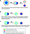
Scheme 1
Scheme 1. Asymmetric Fission of Model Cells
Figure 2
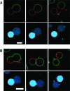
Figure 2
Effect of osmotic stress on two ATPS-containing GVs (A and B) in which lipid membrane phase coexistence was present. The membrane composition for both vesicles was 1:1 DOPC/DPPC + 30% cholesterol, with 2.4% DPPE-PEG-2K, 0.09% DSPE-PEG-2K-biotin, and 0.4% DOPE-rhodamine. Osmolality increases from left to right. Confocoal fluorescence images have been overlaid and false-colored: red is DOPE-rhodamine, indicating the Ld membrane domain, and green is streptavidin-Alexa488, bound to DSPE-PEG-2K-biotin, which is partitioned into the Lo membrane domain. Blue indicates lectin SBA-Alexa 647. Arrows on the far right indicate the location of lipid nanotubes between the daughter vesicles. T = 5 °C. Scale bars are 10 μm.
Figure 3
Figure 3
Confocal fluorescence images collected during asymmetric division of ATPS-containing GV presenting micrometer-scale lipid domains (lipid composition was 1:1 DOPC/DPPC + 30% cholesterol, with 2.2% DPPE-PEG-2K, 0.08% DSPE-PEG2K-carboxyfluorescein, and 0.08% DOPE-rhodamine). Osmolality increases from left to right (130 ± 1.5 mmol/kg to 238 ± 5.5 mmol/kg). Fluorescence images have been false colored: red indicates DOPE-rhodamine in the Ld membrane domain, and green indicates DSPE-PEG 2000-carboxyfluorescein, in the Lo membrane domain, and blue indicates Alexa 647-lectin SBA. The Alexa647 signal decreased over time due to photobleaching; the blue channel has been adjusted to make the partitioning of SBA apparent for each time point. T = 5 °C. Scale bar is 10 μm.
Figure 4
Figure 4
Division of ATPS-containing GVs with excess area of either Ld or Lo membrane domain. Membrane compositions were as follows: 1:1 DOPC/DPPC + 30% cholesterol (A), 1:2 DOPC/DPPC + 30% cholesterol (B). Osmolality increases from left to right. Fluorescence images have been overlaid and false-colored. Blue indicates lectin SBA-Alexa 647, red indicates Ld domain lipid (DOPE-rhodamine), and green indicates Lo domain streptavidin-Alexa488 (bound to lipid DSPE-PEG-2K-biotin). T = 5 °C. Scale bar is 10 μm.
Figure 5
Figure 5
Second-generation aqueous phase separation and budding in a daughter vesicle. Membrane composition was 1:2 DOPC/DPPC + 30% cholesterol. Osmolality increases from left to right. Panels top to bottom are transmitted light (DIC), membrane fluorescence, and interior protein fluorescence. Confocal fluorescence images have been overlaid and false-colored. Red indicates Ld domain lipid (DOPE-rhodamine), green indicates Lo domain lipid (streptavidin-Alexa 488, bound to DSPE-PEG 2000-biotin), and blue indicates the lectin, SBA-Alexa 647, which is partitioned into the dextran-rich interior aqueous phase. T = 5 °C. Scale bar is 10 μm.
Figure 6
Figure 6
Aqueous phase separation in each of resulting vesicles after complete budding to form two daughter vesicles connected by a lipid nanotube. Membrane composition was 1:1 DOPC/DPPC + 30% cholesterol. Osmolality increases from left to right. Top row is transmitted light (DIC). Fluorescence images have been overlaid and false-colored. Green indicates Ld domain lipid (DOPE-CF), red indicates Lo domain (streptavidin-Cy3, bound to lipid DSPE-PEG 2000-biotin), and blue indicates lectin SBA-Alexa 647 (note that the red and green dyes are reversed as compared to previous figures). Arrows highlight the location of newly formed aqueous phases within each of the daughter vesicles. T = 5 °C for the first three panels and 32 °C for the last three panels. Scale bar is 10 μm.

References

    1. Menger F. M.; Gabrielson K. D. Angew. Chem., Int. Ed. Engl. 1995, 34, 2091–2106.
    2. Menger F. M.; Angelova M. I. Acc. Chem. Res. 1998, 31, 789–797.
    3. Oberholzer T.; Luisi P. L. J. Biol. Phys. 2002, 28, 733–744. - PMC - PubMed
    4. Liposomes: A Practical Approach, 2nd ed.; Torchilin V. P., Weissig V., Eds.; Oxford University Press: Oxford, 1990.
    1. Giant Vesicles, Perspectives in Supramolecular Chemistry 6; Luisi P. L., Walde, Eds.; John Wiley and Sons: West Sussex, 2000.
    1. Walde P.; Cosentino K.; Engel H.; Stano P. ChemBioChem 2010, 11, 848–865. - PubMed
    1. Jesorka A.; Orwar O. Annu. Rev. Anal. Chem. 2008, 1, 801–832. - PubMed
    1. Dimova R.; Aranda S.; Bezlyepkina N.; Nikolov V.; Riske K. A.; Lipowsky R. J. Phys.: Condens. Matter 2006, 18, S1151–S1176. - PubMed

Publication types